. 2008 Dec 5;32(5):662-72.
doi: 10.1016/j.molcel.2008.10.022.

Distinct activation pathways confer cyclin-binding specificity on Cdk1 and Cdk2 in human cells

Affiliations

Distinct activation pathways confer cyclin-binding specificity on Cdk1 and Cdk2 in human cells

Karl A Merrick et al. Mol Cell. .

Abstract

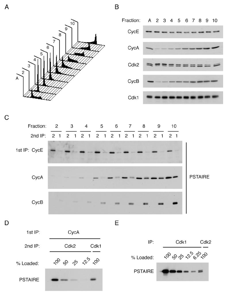
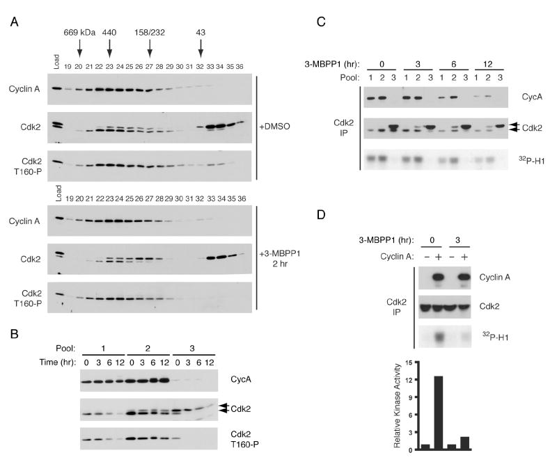
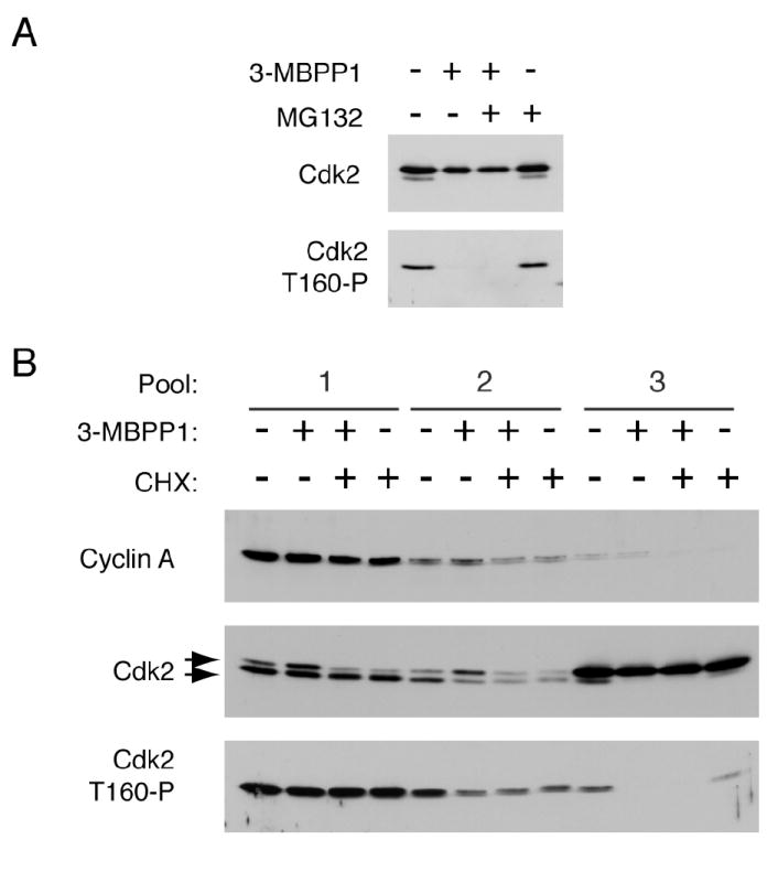
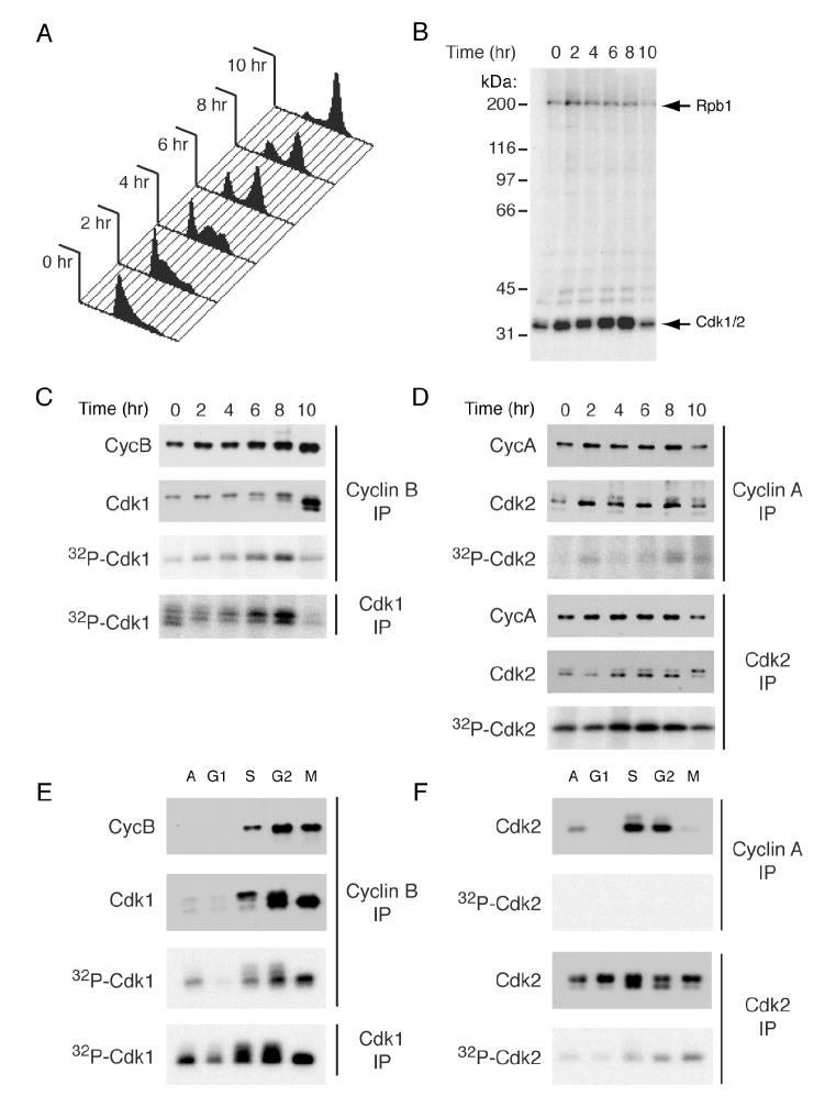
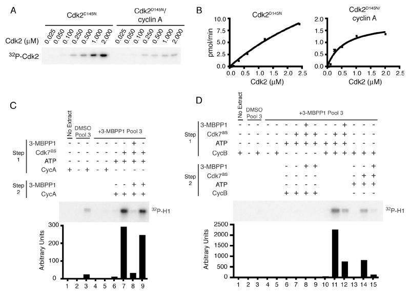
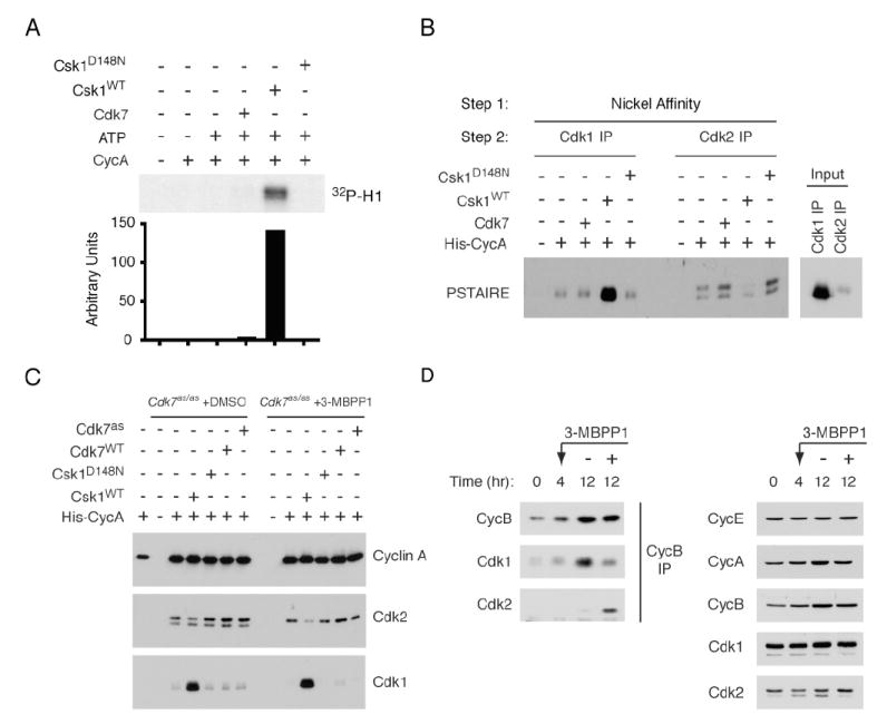
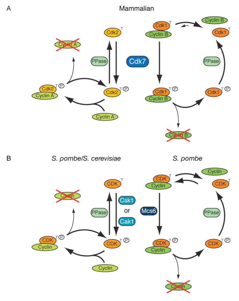
In metazoans, different cyclin-dependent kinases (CDKs) bind preferred cyclin partners to coordinate cell division. Here, we investigate these preferences in human cells and show that cyclin A assembles with Cdk1 only after complex formation with Cdk2 reaches a plateau during late S and G2 phases. To understand the basis for Cdk2's competitive advantage, despite Cdk1's greater abundance, we dissect their activation pathways by chemical genetics. Cdk1 and Cdk2 are activated by kinetically distinct mechanisms, even though they share the same CDK-activating kinase (CAK), Cdk7. We recapitulate cyclin A's selectivity for Cdk2 in extracts and override it with a yeast CAK that phosphorylates monomeric Cdk1, redirecting Cdk1 into a pathway normally restricted to Cdk2. Conversely, upon Cdk7 inhibition in vivo, cyclin B, which normally binds Cdk1 nearly exclusively, is diverted to Cdk2. Therefore, differential ordering of common activation steps promotes CDK-cyclin specificity, with Cdk7 acting catalytically to enforce fidelity.


Figures

Figure 1
Figure 1. Quantitative analysis of CDK/cyclin complex formation
(A) K562 cells were fractionated by centrifugal elutriation and cellular DNA content was determined by flow cytometry. (B) Immunoblots of asynchronous and elutriated K562 cell extracts were probed for cyclins E, A and B, and Cdk1 and -2 (A, asynchronous). (C) Cyclin E, A, or B was immunoprecipitated from whole-cell extracts of elutriated K562 cells, the immunoprecipitates boiled in 1% SDS, the supernatants diluted in HBS + 0.5% Triton-X 100, and Cdk1 and -2 immunoprecipitated. Relative levels of Cdk1 and -2 associated with each cyclin were measured by immunoblotting with PSTAIRE antibody. (D) Cyclin A-bound Cdk1 and -2 were recovered from asynchronous K562 cell extracts and detected as described in C; decreasing amounts of recovered Cdk2 were compared with total Cdk1 recovered to estimate fold-enrichment of Cdk2. (E) Total Cdk1 and -2 in asynchronous cell extract were immunoprecipitated after SDS denaturation, and their relative amounts determined by titrating the anti-Cdk1 eluate against a fixed amount of anti-Cdk2 eluate in a PSTAIRE immunoblot.
Figure 2
Figure 2. Differential Stability of Phospho-Thr160 on Monomeric and Cyclin-Bound Cdk2
(A) Asynchronously grown Cdk7as/as HCT116 cells were treated with DMSO or 5 μM 3-MBPP1 for 2 hr. Extracts were fractionated in a Superdex 200 sizing column, and cyclin A, Cdk2 and T-loop phosphorylated Cdk2 (T160-P) were detected by immunoblot. (B) Pooled sizing column fractions from cells treated for indicated times with 3-MBPP1 were immunoblotted and probed for cyclin A, total Cdk2, and Cdk2 T160-P. Pool 1 contains the highest molecular weight Cdk2, pool 2 the ~200 kDa peak and pool 3 the ~30 kDa peak (see Supplemental Figure 3 for complete data set). The faster-migrating isoform detected by antibodies to total Cdk2 (middle panel, lower arrow) is phosphorylated on Thr160. (C) Cdk2 was immunoprecipitated from pooled fractions in (B) and tested for bound cyclin A by immunoblot (top panel) and associated histone H1 kinase activity (bottom panel). (D) Recombinant His-cyclin A was incubated with pool 3 from DMSO- or 3-MBPP1-treated cells, prior to Cdk2 immunoprecipitation and histone H1 kinase assay. Activity was quantified by phosphorimager.
Figure 3
Figure 3. Thr160 Phosphate Turnover Is Restricted to Cdk2 Monomer In Vivo
(A) Cdk7as/as HCT116 cells were treated with 5 μM 3-MBPP1, 10 μM MG132 or both, as indicated, for 2 hr. Monomeric Cdk2 was isolated by gel exclusion chromatography and probed for Cdk2 and Cdk2 T160-P (see Figure 2A and Supplemental Figure 5 for complete data set). (B) Cdk7as/as HCT116 cells were treated with 5 μM 3-MBPP1, 100 μg/ml cycloheximide or both for 2 hr. Extracts were separated in sizing columns and fractions were pooled (see Supplemental Figure 6 for complete data set). Cyclin A, Cdk2 and Cdk2 T160-P were detected by immunoblot.
Figure 4
Figure 4. Cell-Cycle Dependent Phosphorylation of Cdk7 Substrates in Extracts
(A) Cdk7as/as HCT116 cells were arrested in S phase by addition of thymidine for 15 hr, released into fresh medium containing nocodazole and harvested every 2 hr to determine DNA content by flow cytometry. (B) Extracts from cells treated as in (A) were labeled by endogenous Cdk7as with [γ-32P]N6-(benzyl)-ATP. Only Rpb1 (~200 kDa) and Cdk1 + 2 (~34 kDa), were detected in this exposure. (C) Anti-cyclin B or –Cdk1 immunoprecipitates from labeling reactions in (B) were probed for cyclin B and Cdk1 by immunoblot and for labeling by autoradiography. (D) Anti-cyclin A or -Cdk2 immunoprecipitates from reactions in (B) were probed for cyclin A and Cdk2 by immunoblot and for labeling by autoradiography. (E) Cyclin B and Cdk1 were immunoprecipitated after labeling with purified Cdk7as/cyclin H/Mat1 and [γ-32P]N6-(benzyl)-ATP in extracts of HeLa cells synchronized at indicated cell-cycle positions. Immunoprecipitates were probed for cyclin B and Cdk1 by immunoblot and for phosphorylation by autoradiography. (F) Cyclin A and Cdk2 were immunoprecipitated from labeling reactions as in (E). Cdk2 protein was detected by immunoblot, and the extent of labeling by autoradiography, from single exposures of the same membrane and dried gel, respectively.
Figure 5
Figure 5. Cdk2 but not Cdk1 can be Activated by a CAK-First Pathway
(A) Titrations of Cdk2D145N and Cdk2D145N/cyclin A to determine kinetic parameters of phosphorylation by Cdk7/cyclin H/Mat1 (Supplemental Table 1). The D145N mutation renders Cdk2 inactive, eliminating background due to autophosphorylation. (B) Concentration-velocity plots for reactions in (A). Error bars denote standard deviation of mean in triplicate measurements. (C) Monomeric Cdk2 was immunoprecipitated from pool 3 fractions of DMSO-or 3-MBPP1-treated cells. In lanes 2-5, immobilized Cdk2 was incubated with or without His-cyclin A and tested directly for kinase activity. In lanes 6-9, Cdk2 was first incubated with ATP (all samples), with Cdk7as and 3-MBPP1 added as indicated. After washes, beads were incubated with cyclin A with or without 3-MBPP1, washed again and tested for kinase activity, quantified by phosphorimager. (D) Cdk1 was immunoprecipitated from pool 3 fractions of DMSO- or 3MBPP1-treated cells. In lanes 2-5, immobilized Cdk1 was incubated with purified Myc-cyclin B, washed and tested for kinase activity. In lanes 6-9, Cdk1 was treated exactly as described for lanes 6-9 of (C), except cyclin B was added in place of cyclin A. In lanes 10-12, Cdk1 underwent a single treatment with ATP, Cdk7as, 3-MBPP1 and cyclin B as indicated. In lanes 13-15, Cdk1 was incubated first with cyclin B and, after washing, with ATP, Cdk7as and 3-MBPP1 as indicated. Complexes were then tested for kinase activity, quantified by phosphorimager.
Figure 6
Figure 6. CAKs Influence Specificity of CDK/Cyclin Pairing In Vitro and In Vivo
(A) Cdk1 was immunoprecipitated from pool 3 fractions of 3-MBPP1-treated cells and incubated with purified His-cyclin A without other treatments or, in indicated lanes, after pre-treatment with CAK (Cdk7 or Csk1) and ATP. Beads were washed and tested for histone H1 kinase activity, quantified by phosphorimager. (B) Purified Cdk7 complex, or wild-type (WT) or kinase-dead (D148N) Csk1, were added as indicated to Cdk7as/as HCT116 extracts supplemented with His-cyclin A. After incubation, cyclin A-containing complexes were recovered on nickel beads, denatured and immunoprecipitated with Cdk1-or Cdk2-specific antibodies. In Input lanes, Cdk1 or Cdk2 was immunoprecipitated directly from denatured extract. Recovered CDK was detected with anti-PSTAIRE antibody. (C) Extracts from Cdk7as/as HCT116 cells, treated as indicated with DMSO or 3-MBPP1 for 3 hr, were incubated with His-cyclin A and exogenous CAKs as indicated. Cyclin A-containing complexes were recovered on nickel beads and relative amounts of recovered Cdk1 and -2 determined by immunoblot. (D) Cyclin B was immunoprecipitated from extracts of Cdk7as/as HCT116 cells, synchronized by serum starvation and release and treated at 4 hr with DMSO or 5 μM 3-MBPP1, as indicated, for 8 hr. Associated Cdk1 and-2 were detected by immunoblot (left: anti-Cdk2 blot was exposed ~60-fold longer than was anti-Cdk1 blot). Total levels of Cdk1 and -2, and cyclins A, B, and E were determined by immunoblot (right).
Figure 7
Figure 7. Ordering CDK Activation Pathways in Yeast and Metazoans
(A) In mammalian cells, CDKs follow distinct kinetic pathways to activation. Cdk2 is phosphorylated by Cdk7 before binding cyclin. Cdk7 can only phosphorylate Cdk1 in the presence of cyclin, but Cdk1/cyclin complexes are unstable unless phosphorylated by Cdk7. Upon degradation of its cyclin, Cdk2 can rejoin the monomer pool and be re-phosphorylated, whereas Cdk1 must await association with cyclin for re-phosphorylation. (B) S. pombe could have separate mechanisms of CDK activation analogous to those in mammalian cells, by virtue of having two CAKs with different substrate specificities. The predominant activation pathway in S. cerevisiae, with only one CAK, resembles that of human Cdk2.

References

    1. Aleem E, Kiyokawa H, Kaldis P. Cdc2-cyclin E complexes regulate the G1/S phase transition. Nat Cell Biol. 2005;7:831–836. - PubMed
    1. Arooz T, Yam CH, Siu WY, Lau A, Li KK, Poon RY. On the concentrations of cyclins and cyclin-dependent kinases in extracts of cultured human cells. Biochemistry. 2000;39:9494–9501. - PubMed
    1. Berthet C, Aleem E, Coppola V, Tessarollo L, Kaldis P. Cdk2 knockout mice are viable. Curr Biol. 2003;13:1775–1785. - PubMed
    1. Berthet C, Klarmann KD, Hilton MB, Suh HC, Keller JR, Kiyokawa H, Kaldis P. Combined loss of Cdk2 and Cdk4 results in embryonic lethality and Rb hypophosphorylation. Dev Cell. 2006;10:563–573. - PubMed
    1. Bloom J, Cross FR. Multiple levels of cyclin specificity in cell-cycle control. Nat Rev Mol Cell Biol. 2007;8:149–160. - PubMed

Publication types

LinkOut - more resources