. 2009 Mar 16;178(1-3):250-8.
doi: 10.1016/j.cbi.2008.11.007. Epub 2008 Nov 18.

Posttranslational glutathiolation of aldose reductase (AKR1B1): a possible mechanism of protein recovery from S-nitrosylation

Affiliations

Posttranslational glutathiolation of aldose reductase (AKR1B1): a possible mechanism of protein recovery from S-nitrosylation

Shahid Pervez Baba et al. Chem Biol Interact. .

Abstract

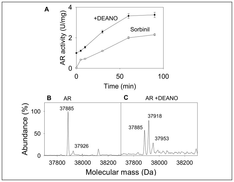
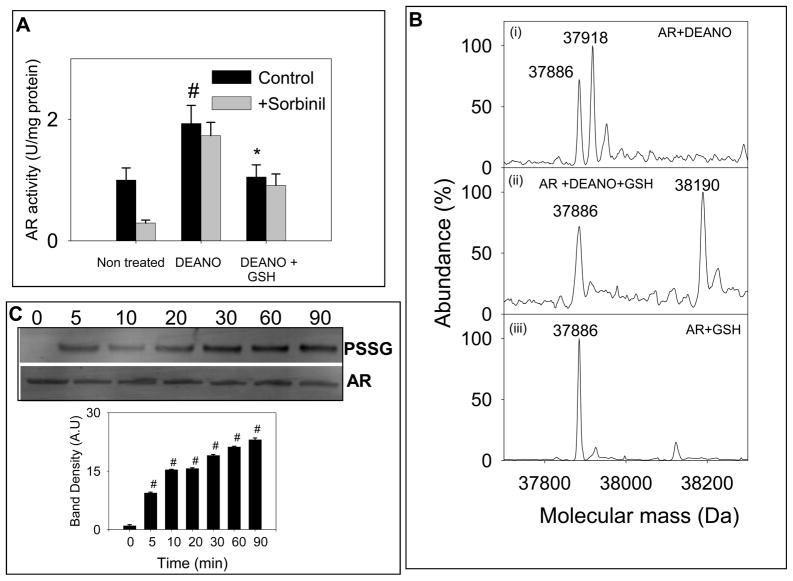
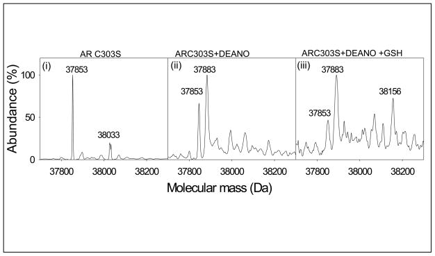
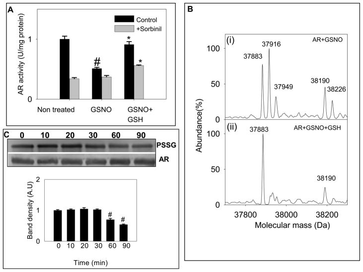
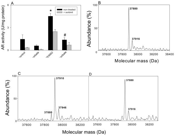
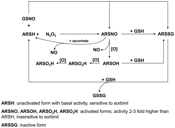
Nitric oxide (NO) is an important regulator of the catalytic activity of aldose reductase (AR). It reacts with the active site cysteines of AR and this reaction results in the formation of several kinetically distinct forms of the protein. The catalytic activity of AR is increased in the ischemic heart and this increase in activity is associated with NO-dependent modification of AR. During reperfusion, the enzyme reverts back to its un-activated form. Although, AR activation has been linked to thiol oxidation, the mechanisms of de-activation remain unclear. Here we report that treatment of recombinant human AR (AKR1B1) by a non-thiol-based NO-donor (DEANO) results in activation and S-nitrosylation of the protein. The nitrosylated (ARSNO), but not the reduced (ARSH), protein reacted with reduced glutathione (GSH) and this reaction resulted in the formation of glutathiolated AR (ARSSG). The modification of AR by NO was site-specific at Cys-298 and was not affected by selective mutation of the neighboring residue, Cys-303 to an alanine. Incubation of the glutathiolated AR (ARSSG) with GSH resulted in the regeneration of the reduced form of the protein (ARSH). Treatment of nitrosylated AR (ARSNO) with ascorbic acid also led to the conversion of the protein to its reduced form. These observations suggest that intracellular reductants such as GSH and ascorbate could convert the nitrosylated form of AR to its basal or reduced state. In general, such reductive reactions might represent a common mechanism for denitrosylating proteins or an "off" switch in NO-mediated signaling pathways involving protein S-nitrosylation reactions.


Figures

Fig. 1
Fig. 1. Modification and activation of AR by NO
(A) Time-dependent changes in the catalytic activity and sorbinil sensitivity of AR. Recombinant human WT His-tag AR was purified from E.coli, reduced with 0.1 M DTT and desalted on a Sephadex G-25 column equilibrated with 0.1 M potassium phosphate (pH 7.0). The reduced enzyme was then incubated with 0.5 mM DEANO at 25°C. Aliquots were withdrawn at indicated times and the catalytic activity was measured with 10 mM DL-glyceraldehyde in the absence (●) and the presence (○) of 1μM sorbinil. Data are shown as discrete points (means ± SD; n=3). (B) Electrospray mass spectrum of WT AR and (C) AR after DEANO treatment. For modification studies the reduced enzyme was incubated with 0.5 mM DEANO in potassium phosphate (pH 7.0) for 60 min at 25 °C. Excess DEANO was removed by passing through Sephadex G-25 column equilibrated with N2-saturated 10 mM ammonium acetate. The deconvoluted electrospray-mass spectrum of unmodified protein corresponds to a single protein with an estimated mass of 37,885 Da, and unidentified minor peak of 37,926 Da. Modification by DEANO resulted in the appearance of protein forms with masses of 37,918 Da and 37,953 Da. The species corresponding to 37,885 Da was assigned to the unmodified protein.
Fig. 2
Fig. 2. Inactivation of S-nitrosylated AR by reduced glutathione
(A) Reduced AR was incubated with 0.5 mM DEANO for 1h at 25°C, desalted and then incubated with 2 mM GSH in 0.1 M potassium phosphate (pH 7.0) for 1h at 25°C. Aliquots were withdrawn from the reaction mixture and the enzyme activity was measured as described under Materials and Methods in the presence and absence of 1 μM sorbinil, as indicated. Values are expressed as mean ± S.D, (n = 3) #P< 0.05 versus non-treated protein and *P <0.05 versus DEANO-treated protein. (B) Electrospray mass spectra of (i) reduced WT AR treated with DEANO showing molecular ions for unmodified AR (37,885 Da) and ARS-NO (37,918 Da); (ii) DEANO-modified AR incubated with 2 mM GSH for 1 h showing both, native (37,885 Da) and glutathiolated (+ 306 Da) AR (38,190 Da); (iii) reduced WT AR incubated with GSH showing only the native protein (37,885 Da). (C) Western blot analysis of DEANO-treated AR incubated with GSH. WT AR was treated with DEANO, desalted and treated with 2 mM GSH for the indicated time periods. The Western blots were developed using monoclonal anti-PSSG antibody. Lower panel shows group data for the intensity of the anti-PSSG immunopositive bands, normalized to AR. Data are presented as mean ± SD. #P < 0.005 versus DEANO-treated protein incubated with GSH for 0 min (n = 3).
Fig. 3
Fig. 3. Identification of the glutathione binding site of AR
Electrospray mass spectrum of (i) AR:C303A (37,853Da) and (ii) AR:C303A incubated with 0.5 mM DEANO in N2-saturated potassium phosphate (0.1 M, pH 7.0) at 25 °C for 60 min. Excess DEANO was removed by passing the reaction mixture through the Sephadex G-25 column and eluted with N2-saturated 10 mM ammonium acetate. Spectra show the native (37,853 Da) and S-nitrosylated protein (37,883 Da); and (iii) AR:C303A incubated with DEANO, desalted and treated with 2 mM GSH. The spectrum shows native (37,853 Da), S-nitrosylated (37,883 Da) and glutathiolated (38,156 Da) forms of the protein.
Fig. 4
Fig. 4. Reduction and reactivation of GSNO modified AR by GSH
(A) Reversal of GSNO-induced inhibition of the enzyme by GSH. The reduced enzyme was incubated with 50 μM GSNO in 0.1 M potassium phosphate (pH 7.0) for 1 h at 25°C. The treated protein was desalted and incubated with 2 mM GSH for 1 h at 25°C. At the end of the treatment aliquots were withdrawn and assayed for enzyme activity without and with 1 M sorbinil, as indicated. Values are expressed as mean ± S.D, (n = 3) # P< 0.05 versus non-treated protein and * P < 0.05 versus GSNO-treated protein. Electrospray mass spectra of AR (i) incubated with 50 μM GSNO or 1 h at room temperature showing major species corresponding to native (37,883 Da), S-nitrosylated (37,916 Da) and glutathiolated (38,190 Da) forms of the protein and (ii) treated with GSNO, desalted and incubated with 2 mM GSH for 1 h at room temperature. The spectrum shows a major species corresponding to the native (37,883 Da) and a minor peak corresponding to a glutathiolated (38,190 Da) form of the protein. (C) Western blot analysis of time-dependent changes in glutathiolated AR incubated with 2 mM GSH. AR was treated with GSNO and incubated with GSH for the indicated time periods. PVDF membrane was immunoblotted by using anti-PSSG antibody. Lower panel shows group data for the intensity of the anti-PSSG immunopositive bands normalized to AR. Data are expressed as Mean ± SD. #P < 0.005 versus GSNO-treated protein incubated with GSH for 0 min (n = 3).
Fig. 5
Fig. 5. De-activation and de-nitrosylation of AR by ascorbic acid
(A) Catalytic activity of reduced and S-nitrosylated WT AR treated with ascorbic acid. Aliquots of reduced AR were treated with 0.5 mM DEANO alone for 1 h, or treated with DEANO followed by the incubation with 10 mM sodium ascorbate for 1 h, as indicated. Prior to enzyme activity measurements, salts were removed by the gel filtration and AR activity was measured as described under Materials and Methods. Sensitivity of the modified enzyme to 1 μM sorbinil was measured in parallel. Data are reported as mean ± SD (n=3). *P < 0.01 versus control; #P < 0.02 versus DEANO. Electrospray mass spectra of (B) native AR (37,880) (C) native AR treated with 0.5 mM DEANO showing the addition of one (37,910 Da) NO molecule bound to the protein; and (D) AR treated with 0.5 mM DEANO followed by the incubation with 10 mM sodium ascorbate, showing only the reduced native form of the AR protein (37,880 Da).
Scheme I
Scheme I. Modification of AR by NO and nitrosoglutathione (GSNO)
Reduced AR (ARSH) is nitrosylated by N2O3 (generated from the reaction of NO with molecular oxygen) to the activated (ARSNO) form. Reaction of AR with GSNO results mainly in the formation of glutathiolated AR (ARSSG). Nitrosylated AR (ASNO) is generated as a minor side product. The ARSSG form is catalytically inactive. The nitrosylated form of AR (ARSNO) could be oxidized and de-nitrosylated to generate oxy-sulfur acid forms of AR (ARSOH, ARSO2H, and ARSO3H). Like ARSNO, these forms of AR display 2–3 fold higher catalytic activity and are relatively insensitive to inhibition by sorbinil. In addition to oxidation, ARSNO could react with GSH to generate ARSSG which is catalytically inactive. Although not shown in the current study, ARSOH could also be converted to ARSSG by GSH. The ARSSG form is subsequently reduced to the basal ARSH form by another GSH molecule. ARSNO could also be reduced and converted back to ARSH by ascorbic acid. The catalytic activity of each of these forms is listed. Several forms of AR could, therefore, coexist in a cell depending upon the concentration of GSH, NO, and oxygen.

References

    1. Srivastava SK, Ramana KV, Bhatnagar A. Role of aldose reductase and oxidative damage in diabetes and the consequent potential for therapeutic options. Endocr Rev. 2005;26:380–392. - PubMed
    1. Nakayama M, Nakamura J, Hamada J, Chaya S, Mizubayashi R, Yasuda Y, Kamiya K, Koh N, Hotta N. Aldose reductase inhibition ameliorates pupillary light reflex and F-wave latency in patients with mild diabetic neuropathy. Diabetes Care. 2001;24:1093–1098. - PubMed
    1. Hodgkinson AD, Sondergaard KL, Yang BM, Cross DF, Millward BA, Demaine AG. Aldose reductase expression is induced by hyperglycemia in diabetic nephropathy. Kidney Int. 2001;60:211–218. - PubMed
    1. Chung SSM, Chung SK. Aldose reductase in diabetic microvascular complications. Curr Drug Targets. 2005;6:475–486. - PubMed
    1. Kaiserova K, Srivastava S, Hoetker JD, Awe SO, Tang XL, Cai J, Bhatnagar A. Redox activation of aldose reductase in the ischemic heart. J Biol Chem. 2006;281:15110–15120. - PubMed

Publication types

MeSH terms