. 2009 Jan 30;385(4):1127-41.
doi: 10.1016/j.jmb.2008.11.022. Epub 2008 Nov 24.

Structure of amantadine-bound M2 transmembrane peptide of influenza A in lipid bilayers from magic-angle-spinning solid-state NMR: the role of Ser31 in amantadine binding

Affiliations

Structure of amantadine-bound M2 transmembrane peptide of influenza A in lipid bilayers from magic-angle-spinning solid-state NMR: the role of Ser31 in amantadine binding

Sarah D Cady et al. J Mol Biol. .

Abstract

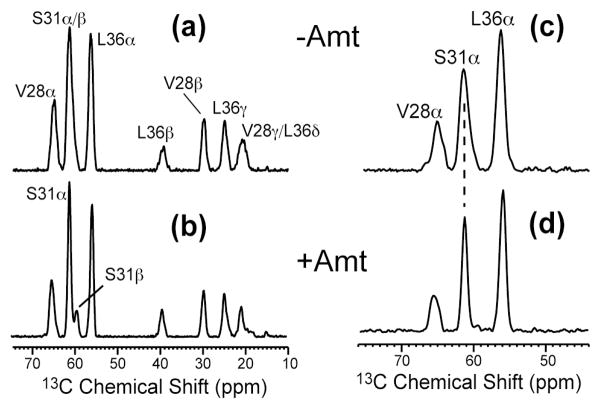
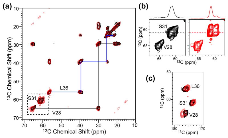
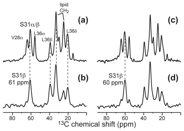
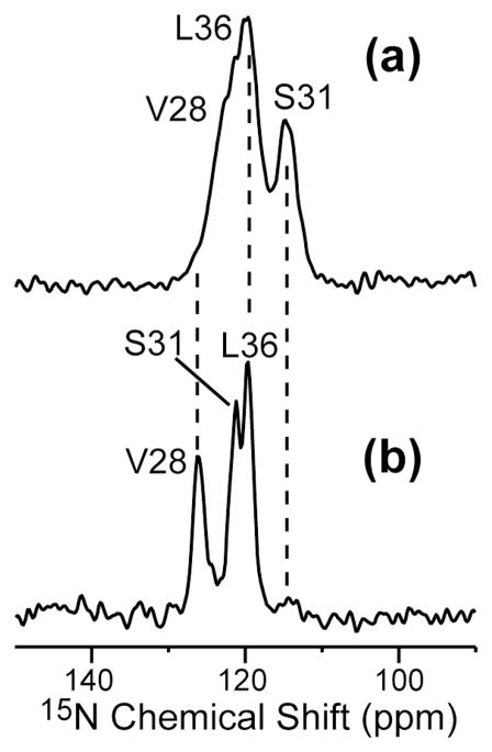
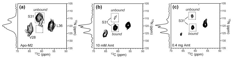
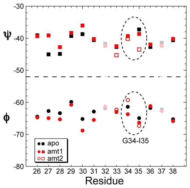
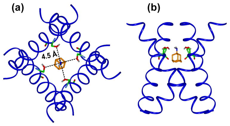
The M2 proton channel of influenza A is the target of the antiviral drugs amantadine and rimantadine, whose effectiveness has been abolished by a single-site mutation of Ser31 to Asn in the transmembrane domain of the protein. Recent high-resolution structures of the M2 transmembrane domain obtained from detergent-solubilized protein in solution and crystal environments gave conflicting drug binding sites. We present magic-angle-spinning solid-state NMR results of Ser31 and a number of other residues in the M2 transmembrane peptide (M2TMP) bound to lipid bilayers. Comparison of the spectra of the membrane-bound apo and complexed M2TMP indicates that Ser31 is the site of the largest chemical shift perturbation by amantadine. The chemical shift constraints lead to a monomer structure with a small kink of the helical axis at Gly34. A tetramer model is then constructed using the helix tilt angle and several interhelical distances previously measured on unoriented bilayer samples. This tetramer model differs from the solution and crystal structures in terms of the openness of the N-terminus of the channel, the constriction at Ser31, and the side-chain conformations of Trp41, a residue important for channel gating. Moreover, the tetramer model suggests that Ser31 may interact with amantadine amine via hydrogen bonding. While the apo and drug-bound M2TMP have similar average structures, the complexed peptide has much narrower linewidths at physiological temperature, indicating drug-induced changes of the protein dynamics in the membrane. Further, at low temperature, several residues show narrower lines in the complexed peptide than the apo peptide, indicating that amantadine binding reduces the conformational heterogeneity of specific residues. The differences of the current solid-state NMR structure of the bilayer-bound M2TMP from the detergent-based M2 structures suggest that the M2 conformation is sensitive to the environment, and care must be taken when interpreting structural findings from non-bilayer samples.


Figures

Figure 1
Figure 1
M2(22–46) sequence and its representation as a heptad repeat. (a) Amino acid sequence. The a and d residues of the heptad repeat abcdefg are labeled. The 13C, 15N-labeled residues that have been studied so far are colored in red. H37 and W41, which are central for proton conduction and channel gating, are shown in blue. (b) Four-helix bundle organization of M2TMP. Channel-facing a and d residues are shaded in pink, and interfacial e and g residues are shaded in cyan.
Figure 2
Figure 2
1D 13C spectra of VSL-M2TMP in DLPC bilayers at 243 K. (a) 13C DQF spectrum of the apo peptide. (b) 13C DQF spectrum of the amantadine-bound peptide. (c) 15N-filtered 13C spectrum of the apo peptide. (d) 15N-filtered 13C spectrum of the amantadine-bound peptide.
Figure 3
Figure 3
2D 13C-13C DQF correlation spectra of VSL-M2TMP in DLPC bilayers at 243 K without (black) and with (red) amantadine. (a) Aliphatic region. (b) S31 and V28 region. (c) Carbonyl region.
Figure 4
Figure 4
Selective detection of S31 Cβ by CH2 editing. (a) 13C CP-MAS spectrum of apo-M2TMP. (b) CH2 edited spectrum of apo-M2TMP. (c) 13C CP-MAS spectrum of amantadine-bound M2TMP. (d) CH2 edited spectrum of bound M2TMP. Note the suppression of the Cα CH peaks in (b) and (d).
Figure 5
Figure 5
1D 15N CP-MAS spectra of VSL-M2TMP in DLPC bilayers at 243 K. (a) Without amantadine. (b) With amantadine.
Figure 6
Figure 6
(a) 2D 15N-13C 2D correlation spectrum of VSL-M2TMP in DLPC bilayers at 243 K. Black: without amantadine. Red: with amantadine. (b) S31 15N cross sections.
Figure 7
Figure 7
Amantadine-induced changes of M2TMP structure and dynamics in DLPC bilayers. (a) Average isotropic chemical shift changes for each residue, measured at low temperature. (b) Average 13C T2 changes, measured at 303 K.
Figure 8
Figure 8
1D 13C variable-temperature CP-MAS spectra of VSL-M2TMP bound to DLPC bilayers. (a) Without amantadine. (b) With amantadine. Temperatures from top to bottom are 313 K, 283 K and 243 K.
Figure 9
Figure 9
2D 15N-13C correlation spectra of VSL-M2TMP in DLPC bilayers with different amounts of amantadine. The S31 15N-13Cα cross peak is used to indicate the degree of amantadine binding. 1D 15N CP spectra are shown projected from the indirect dimension of each spectrum. (a) No amantadine. The S31 peak is completely in the unbound position. (b) Amantadine added at 10 mM in the buffer. The M2 : amantadine molar ratio in the final membrane is ~1 : 8. Almost all S31 intensities are at the bound position. (c) Amantadine directly titrated into the membrane pellet. The M2 : amantadine molar ratio in the final pellet is ~ 1 : 2. 70% of the S31 intensity is at the bound position.
Figure 10
Figure 10
Fractional 13C T2 changes of DLPC lipid carbons upon amantadine binding at 313 K. No M2 peptide is present. The fractional change is calculated as (T2,amt-T2,apo)/T2,apo.
Figure 11
Figure 11
TALOS predicted (ϕ, ψ) angles for residues 26–38 of apo (black) and amantadine-complexed M2TMP (red). Filled and open red symbols denote the conformations of amt1 and amt2, which differ at G34. Residues 32 and 37 are shaded due to incomplete experimental data.
Figure 12
Figure 12
Chemical shift constrained backbone structure of the M2TMP monomer. Apo: black and gray ribbon. Amantadine-bound: blue and cyan ribbon. (a) Side view of the helix with tilt angles determined by SSNMR. (b) Top view of the apo monomer. (c) Top view of the amantadine-complexed monomer. The apo peptide has a more noticeable kink.
Figure 13
Figure 13
Tetramer model of M2TMP constrained by SSNMR chemical shifts, interhelical distances and helix orientations. (a) Trp41 5-19F distance between the two opposite helices is 16.7 Å . (b) Phe30 4-19F diagonal distance is 12.5 Å . (c) Side view of the tetramer, with a diagonal Gly34 Cα-Cα distance of 10.4 Å.
Figure 14
Figure 14
Comparison of the MAS NMR structure model with other recently published structures of drug-complexed M2. The Trp41 indole ring and the diagonal distance between its 5-19F or Hζ3 is indicated. (a) MAS-NMR model of amantadine-bound M2TMP. (a) 15N static SSNMR model of amantadine-bound M2TMP (PDB code: 2H95). (b) Solution NMR structure of rimantadine-bound M2(18-60) (PDB code: 2RLF). (c) Crystal structure of amantadine-bound M2TMP (PDB: 3C9J).
Figure 15
Figure 15
MAS-NMR M2 tetramer model with the proposed amantadine-binding site. The height of amantadine is set to be similar to that of the crystal structure. Ser31 sidechain O distances to the amine are indicated. The top view looks down the helix axis from the N- to the C-terminus.

References

    1. Pinto LH, Lamb RA. Controlling influenza virus replication by inhibiting its proton channel. Mol Biosyst. 2007;3:18–23. - PubMed
    1. Bright RA, Medina MJ, Xu X, Perez-Oronoz G, Wallis TR, Davis XM, Povinelli L, Cox NJ, Klimov AI. Incidence of adamantane resistance among influenza A (H3N2) viruses isolated worldwide from 1994 to 2005: a cause for concern. Lancet. 2005;366:1175–1181. - PubMed
    1. Lamb RA, Holsinger KJ, Pinto LH. The Influenza A virus M2 ion channel protein and its role in the influenza virus life cycle. In: Wemmer E, editor. Cellular Receptors of Animal Viruses. Cold Spring Harbor Lab Press; Plainview, NY: 1994. pp. 303–321.
    1. Pinto LH, Holsinger LJ, Lamb RA. Influenza virus M2 protein has ion channel activity. Cell. 1992;69:517–528. - PubMed
    1. Holsinger LJ, Nichani D, Pinto LH, Lamb RA. Influenza A virus M2 ion channel protein: a structure-function analysis. J Virol. 1994;68:1551–1563. - PMC - PubMed

Publication types

LinkOut - more resources