. 2008 Dec 23;18(24):1978-85.
doi: 10.1016/j.cub.2008.11.023. Epub 2008 Dec 8.

Extracellular sugar modifications provide instructive and cell-specific information for axon-guidance choices

Affiliations

Extracellular sugar modifications provide instructive and cell-specific information for axon-guidance choices

Hannes E Bülow et al. Curr Biol. .

Abstract

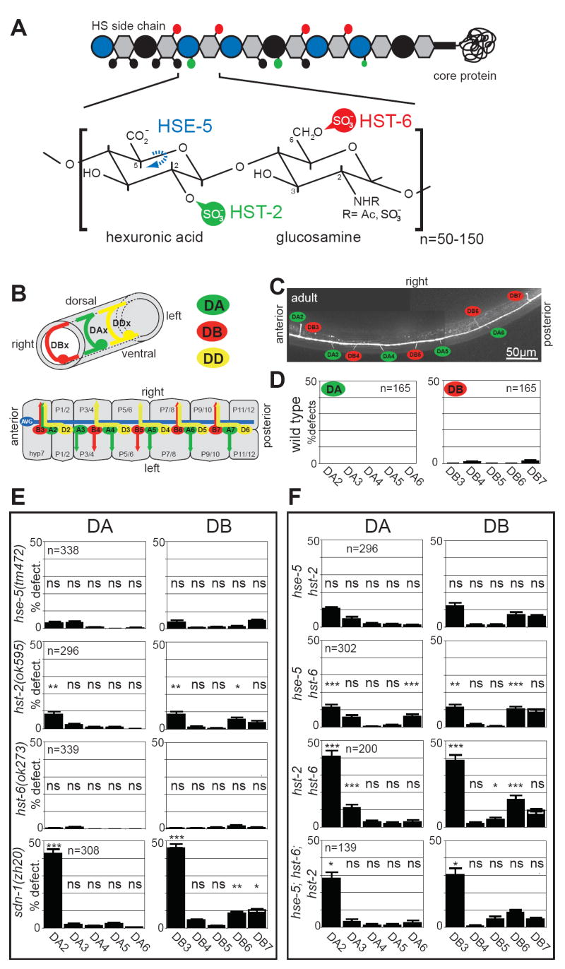
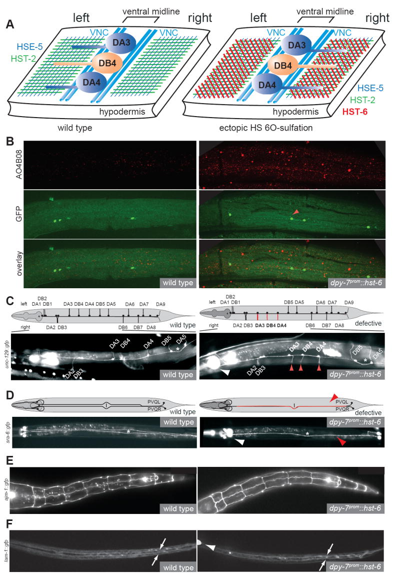
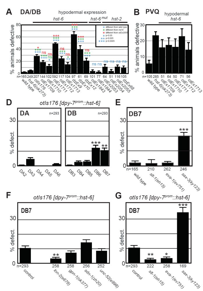
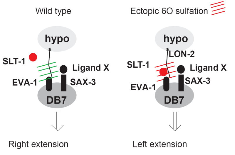
Heparan sulfates (HSs) are extraordinarily complex extracellular sugar molecules that are critical components of multiple signaling systems controlling neuronal development. The molecular complexity of HSs arises through a series of specific modifications, including sulfations of sugar residues and epimerizations of their glucuronic acid moieties. The modifications are introduced nonuniformly along protein-attached HS polysaccharide chains by specific enzymes. Genetic analysis has demonstrated the importance of specific HS-modification patterns for correct neuronal development. However, it remains unclear whether HS modifications provide a merely permissive substrate or whether they provide instructive patterning information during development. We show here with single-cell resolution that highly stereotyped motor axon projections in C. elegans depend on specific HS-modification patterns. By manipulating extracellular HS-modification patterns, we can cell specifically reroute axons, indicating that HS modifications are instructive. This axonal rerouting is dependent on the HS core protein lon-2/glypican and both the axon guidance cue slt-1/Slit and its receptor eva-1. These observations suggest that a changed sugar environment instructs slt-1/Slit-dependent signaling via eva-1 to redirect axons. Our experiments provide genetic in vivo evidence for the "HS code" hypothesis which posits that specific combinations of HS modifications provide specific and instructive information to mediate the specificity of ligand/receptor interactions.


Figures

Fig.1
Fig.1. Individual DA/DB motor neurons require distinct combinations of HS modifications
A: Heparan sulfate is a polymer composed of highly modified disaccharide repeats that is attached to a core protein. Shown is a characteristic HS disaccacharide repeat unit consisting of a hexuronic acid (circle) and a glucosamine (hexagon) residue and enzymes that modify this structure. Large blue vs. black circles indicate iduronic acid vs. glucuronic acid, respectively. Small circles in black, green and, red indicate modifications as color coded in the disaccharide repeat. HSE-5: HS C5 epimerase; HST-X: HS XO-sulfotransferase (X=2, 6). B: The upper panel shows the circumferential axonal trajectory of exemplary DA, DB and DD-type eMNs. eMNs are distinguished by their axonal projection patterns and by the neurotransmitter, which they express. The lower panel shows a schematic “open book” representation of the 2- to 3-fold embryonic stage when eMN axon outgrowth occurs, illustrating that individual DA, DB eMNs show a distinct sidedness of circumferential axonal growth. The eMN cell bodies shown here lie on top of the junctions between bilaterally symmetry P1/2, P3/4, P5/6, P7/8, P9/10 and P11/12 hypodermal cells. More anteriorly and posteriorly positioned eMNs are not shown (see Experimental Procedures). The only other axon that populates the VNC at that stage is the axon of the AVG pioneer neuron. See Supplementary Table 1 for a systematic comparison of individual motor neuron class members. C: Visualizing the DA and DB motor axons with an unc-129∷gfp expressing transgene. D: The left/right asymmetric, circumferential DA, DB axon projection patterns are highly stereotyped. We focus our scoring on the DA2 to DA6 and DB3 to DB7 motor neurons since those express the gfp reporter transgene most reliably. “% defect.” indicates percentage of animals with a defective sidedness of the respective DA or DB axon as indicated. E: Loss of HS modifications affect DA and DB axon choices in a cell-type specific manner. All mutants used are null mutants [7, 30]. Single mutants were statistically compared to wild type (see D.); ns = not significantly different; * P<0.05, ** P<0.005. F: Comparison of effects of single, double and, triple HS mutants on specific motor axons. Double mutants were statistically compared to the more severe of two respective single mutants; ns = not significantly different; * P<0.05, ** P<0.005, *** P<0.0005. The defects in both DA2 and DB3 are weakly statistically significant when comparing the triple hst-2 hst-6 hse-5 mutant and the syndecan null mutant (p=0.04 and 0.03, respectively). However, these differences are not significant between the hst-2 hst-6 double mutant and either the sdn-1 mutant or the hst-2 hst-6 hse-5 triple mutant suggesting that the phenotype of the sdn-1 and the hst-2 hst-6 hse-5 triple mutant is very similar. See methods for statistics.
Fig.2
Fig.2. Manipulating HS modification patterns redirects motor axons
A: Schematic view of the ventral cord with HS modifications in the hypodermis indicated by colored hatchings. Note that unlike in larvae, the embryonic midline, defined as a physical barrier between the left and right ventral fascicles, is not constituted by a hypodermal evagination but by motor neuron cell bodies [31]. B: Immunostaining with the AO4B08 HS specific antibody [15] of transgenic animals, which ectopically express the HS modification enzyme hst-6 in the hypodermis (right panels; otIs176). Increased hypodermal staining observed in transgenic animals. This increased staining is not seen in isogenic controls, which do not ectopically express hst-6 (left panels). Intestinal staining (not shown) serves as an internal staining control as does an unrelated synthetic antibody (MPB49), which shows no staining at all. Note that the antibody AO4B08 does not recognize individual HS modifications but rather an oligosaccharide, which requires the activities of both, hst-2 and hst-6 [15]. Since only hst-2 but not hst-6 appears to be expressed in the hypodermis [7], the observed staining of the hypodermis following misexpression of hst-6 in this tissue are consistent with the known specificities of AO4B08. Note the aberrant projection of DB6 in the hst-6 misexpressing line (red arrowhead). C: Axonal misrouting of DA/DB motor neurons in animals with ectopic hst-6 expression in the hypodermis (right panel, otEx1711). Dashed lines indicate neurons that were not scored and, affected neurons are in red. See Fig.3 and Suppl. Fig.3 for quantification of defects. D: Midline crossover defects of PVQ interneurons in animals with ectopic hst-6 expression in the hypodermis (right panel, otEx1711). E: Hypodermal cell fate as well as cell morphology, as visualized by normal AJM-1 expression and localization patterns, is not visibly affected by ectopic hst-6 expression (right panel, otIs176). F: Basement membrane topology, as visualized by a lam-1∷gfp reporter (kind gift of David Sherwood)[32], is not visibly affected by ectopic hst-6 expression (right panel, otIs176). Arrows point to the basement membrane that ensheathes the ventral midline, and which visually appears most concentrated on each side of the midline as a result of optical sectioning. Left panels represent wild type controls in all cases. White arrowheads point to pharyngeal expression of gfp denoting the presence of the hypodermal hst-6 misexpression array and red arrowheads indicate axonal routing errors. All views are ventral aspects with anterior to the left.
Fig.3
Fig.3. Genetic analyses of hst-6-dependent axonal misrouting phenotypes
A: Quantification of DA/DB motor neuron defects in different transgenic strains that express the indicated constructs from the hypodermal specific dpy-7 promoter. Shown is the percentage of animals with any number (≥1 per animal) of DA or DB axon sidedness defects. B: Quantification of midline cross over defects in PVQ interneurons in different transgenic strains that express hst-6 from the hypodermal specific dpy-7 promoter. C: Effect of ectopic hst-6 expression on individual DA and DB neurons. One representative line is shown here, 5 more extrachromosomal lines are shown in Suppl. Fig.2B. Shown is the percentage of animals with defects in individual DA/DB motor axons projections as indicated. D: Defects in DB7 motor axon projection patterns in different mutants of the Slit/Robo signaling pathway as indicated. Shown is the percentage of animals with defects in DB7 motor axon projections. E: Effect of genetic removal of HS core proteins on projection errors in DB7 motor axons as a result of ectopic hst-6 expression. Shown is the percentage of animals with defects in DB7 motor neuron projections. Statistic comparisons are made to the transgenic control. The projection pattern of DB7 in animals that misexpress hst-6 in the hypodermis and are also mutant for lon-2, is statistically indistinguishable from non-transgenic, wild-type animals. F: Effect of removal of genes in the Slit/Robo signaling pathway on projection errors in DB7 motor axons as a result of ectopic hst-6 expression. Shown is the percentage of animals with defects in DB7 motor axon projections. Statistic comparisons are made to the transgenic control. The projection pattern of DB7 in animals that misexpress hst-6 in the hypodermis and are also mutant for slt-1 or eva-1, is statistically indistinguishable from non-transgenic, wild-type animals. Statistical significance is indicated by: ns, not significantly different, * P<0.05, ** P<0.005, *** P<0.0005 in all panels.
Fig.4
Fig.4. A model for the role of HS in redirecting axonal projections
A possible interpretation of the genetic data presented in Fig.3. In wild-type animals, slt-1 or eva-1 is not required for DB7 neuron projections (as slt-1 and eva-1 mutants show no axonal defects; Fig.3D), while sax-3 does normally have a role in DB7 axon extension (Fig.3D). In wild-type animals, SAX-3 may therefore be interacting with an as yet unknown ligand X. Upon introducing ectopic 6O-sulfation patterns (red hatches), an interaction of the SLT-1 protein with EVA-1 may be promoted, which supersedes the effect normally induced by SAX-3/Ligand X. We emphasize that these conclusions are based on purely genetic arguments and that more indirect mechanistic models can be envisioned which are not shown here.

Similar articles

Cited by

References

    1. Lee JS, Chien CB. When sugars guide axons: insights from heparan sulphate proteoglycan mutants. Nat Rev Genet. 2004;5:923–935. - PubMed
    1. Van Vactor D, Wall DP, Johnson KG. Heparan sulfate proteoglycans and the emergence of neuronal connectivity. Curr Opin Neurobiol. 2006;16:40–51. - PubMed
    1. Bülow HE, Hobert O. The Molecular Diversity of Glycosaminoglycans Shapes Animal Development. Annu Rev Cell Dev Biol 2006 - PubMed
    1. Esko JD, Selleck SB. Order out of chaos: assembly of ligand binding sites in heparan sulfate. Annu Rev Biochem. 2002;71:435–471. - PubMed
    1. Turnbull J, Powell A, Guimond S. Heparan sulfate: decoding a dynamic multifunctional cell regulator. Trends Cell Biol. 2001;11:75–82. - PubMed

Publication types

MeSH terms