Review
. 2009 Mar;15(1):29-41.
doi: 10.1089/ten.teb.2008.0329.

Morphogenetic and regulatory mechanisms during developmental chondrogenesis: new paradigms for cartilage tissue engineering

Affiliations
Review

Morphogenetic and regulatory mechanisms during developmental chondrogenesis: new paradigms for cartilage tissue engineering

Lluís Quintana et al. Tissue Eng Part B Rev. 2009 Mar.

Abstract

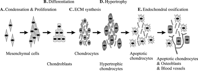
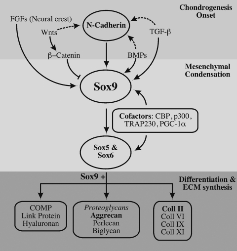
Cartilage is the first skeletal tissue to be formed during embryogenesis leading to the creation of all mature cartilages and bones, with the exception of the flat bones in the skull. Therefore, errors occurring during the process of chondrogenesis, the formation of cartilage, often lead to severe skeletal malformations such as dysplasias. There are hundreds of skeletal dysplasias, and the molecular genetic etiology of some remains more elusive than of others. Many efforts have aimed at understanding the morphogenetic event of chondrogenesis in normal individuals, of which the main morphogenetic and regulatory mechanisms will be reviewed here. For instance, many signaling molecules that guide chondrogenesis--for example, transforming growth factor-beta, bone morphogenetic proteins, fibroblast growth factors, and Wnts, as well as transcriptional regulators such as the Sox family--have already been identified. Moreover, extracellular matrix components also play an important role in this developmental event, as evidenced by the promotion of the chondrogenic potential of chondroprogenitor cells caused by collagen II and proteoglycans like versican. The growing evidence of the elements that control chondrogenesis and the increasing number of different sources of progenitor cells will, hopefully, help to create tissue engineering platforms that could overcome many developmental or degenerative diseases associated with cartilage defects.


Figures

FIG. 1.
FIG. 1.
Schematic representation of the chondrogenesis and endochondral ossification. (A) First MCs condense to form a dense cell mass. (B) MCs proliferate and differentiate into chondroblasts. (C) These cells start secreting cartilage ECM and become mature chondrocytes. (D) Eventually, chondrocytes grow to become hypertrophic, and if the tissue undergoes endochondral ossification, (E) cartilage is vascularized, ECM is degraded, hypertrophic chondrocytes become apoptotic, and osteoblasts invade the free space within the tissue.
FIG. 2.
FIG. 2.
Upstream and downstream regulation of Sox9. Chondrogenesis begins with the upregulation of N-cadherin and Sox9 by paracrine factors like TGF-β, FGFs, or BMPs, while Wnts downregulate the expression of Sox9. Expression of N-cadherins and Sox9 is accompanied by the condensation and proliferation of MCs, a process called mesenchymal condensation. Sox9 activates the expression of Sox5 and Sox6, and with them and the help of cofactors such as CBP, induces the MC to differentiate into chondroblasts after the condensation. Chondroblasts produce cartilage-specific ECM, including collagen II, IV, IX, and XI; proteoglycans such as aggrecan; and also link proteins and COMP.
FIG. 3.
FIG. 3.
In vitro models for chondrogenesis and osteogenesis with embryonic primitive cells (mESCs and mouse embryonic fibroblasts [MEFs]). (A, B) Chondrocytes were derived from mESCs with BMP-2 and TGF-β1 as described. After 4 weeks of in vitro culture, cartilage nodules appear that contain small round cells (B) that secrete a grayish matrix that differs from mature mineralized osteogenic cultures shown in (C). (D) Toluidine blue staining of MEFs cultured in RAD16-I with Dulbecco's modified Eagle's medium (DMEM) supplemented with fetal bovine serum (FBS), L-Glu, and antibiotics reveals that these cells spontaneously undergo chondrogenesis and produce proteoglycans. (E) Von Kossa staining of MEFs after 21 days cultured in RAD16-I with DMEM supplemented with FBS, L-Glu, β-glycerophosphate, dexamethasone, sodium ascorbate, and antibiotics. MEFs can also promote calcium deposition and become osteoblast-like cells. (F) mESCs after osteogenic induction in RAD16-I hydrogels produce osteoblasts-like cells that induce calcium salts deposition, indicated by white arrows. Scale bars: 100 μm. Color images available online at www.liebertonline.com/ten.
FIG. 4.
FIG. 4.
Endochondral bone formation from mESCs in vitro. mESCs were differentiated with 1,25-OH2 vitamin D3 toward osteoblasts, which appear black in phase contrast microscopy. Chondrogenic cultures were treated with BMP-2 from day 3 of differentiation onward. Interestingly, chondrogenic cultures can undergo mineralization when additionally triggered with the osteogenic inducer 1,25-OH2 vitamin D3. However, a window of opportunity seems to exist that falls between day 15 and day 20 of differentiation.

Similar articles

Cited by

References

    1. Newman S.A. Frisch H.L. Dynamics of skeletal pattern formation in developing chick limb. Science. 1979;205:662. - PubMed
    1. Horton W.A. Hood O.J. Machado M.A. Ahmed S. Griffey E.S. Abnormal ossification in thanatophoric dysplasia. Bone. 1988;9:53. - PubMed
    1. Hall B.K. Miyake T. Divide, accumulate, differentiate: cell condensation in skeletal development revisited. Int J Dev Biol. 1995;39:881. - PubMed
    1. Hoffman L.M. Weston A.D. Underhill T.M. Molecular mechanisms regulating chondroblast differentiation. J Bone Joint Surg Am. 2003;85-A Suppl 2:124. - PubMed
    1. Mackie E.J. Ahmed Y.A. Tatarczuch L. Chen K.S. Mirams M. Endochondral ossification: how cartilage is converted into bone in the developing skeleton. Int J Biochem Cell Biol. 2008;40:46. - PubMed

Publication types