

Scrotal calcinosis due to resorption of cyst walls: a case report
- PMID: 19063719
- PMCID: PMC2629479
- DOI: 10.1186/1752-1947-2-375
Scrotal calcinosis due to resorption of cyst walls: a case report
Abstract
Introduction: Scrotal calcinosis is a rare benign entity defined as the presence of multiple calcified nodules within the scrotal skin. There are controversies about the origin of this entity. In fact, it is still debatable whether scrotal calcinosis is an idiopathic growth or dystrophic calcification of dartoic muscles. It is also unclear whether scrotal calcinosis originates from inflammation of epidermal cysts affected by mild to moderate inflammation of mononuclear cells, from foreign body granuloma formation followed by resorption of cyst walls or from eccrine epithelial cysts.
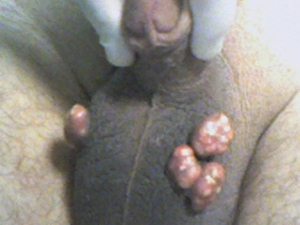
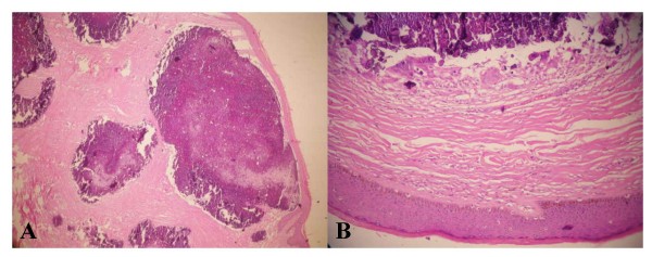
Case presentation: We report a 41-year-old male Turkish patient presenting with a 10-year history of scrotal tumours increasing slowly in size and number. Histopathologically, there was no epithelial lining around the calcified nodules, but there was fibrosis adjacent to atrophic stratified squamous epithelium.
Conclusion: Results of histopathological examinations suggested that scrotal calcinosis might have been due to resorption of cyst walls. Surgery remains the key for this problem. In cases of non-massive scrotal calcinosis, like the case presented here, excision of the nodules from the affected part of the scrotal wall and repairing the defect with horizontal stitches offer good cosmetic results without relapse.
Figures
References
LinkOut - more resources
Full Text Sources
