. 2009 Feb 10;184(3):186-91.
doi: 10.1016/j.toxlet.2008.11.008. Epub 2008 Nov 21.

Analysis of targeted mutation in DJ-1 on cellular function in primary astrocytes

Affiliations

Analysis of targeted mutation in DJ-1 on cellular function in primary astrocytes

Amanda K Ashley et al. Toxicol Lett. .

Abstract

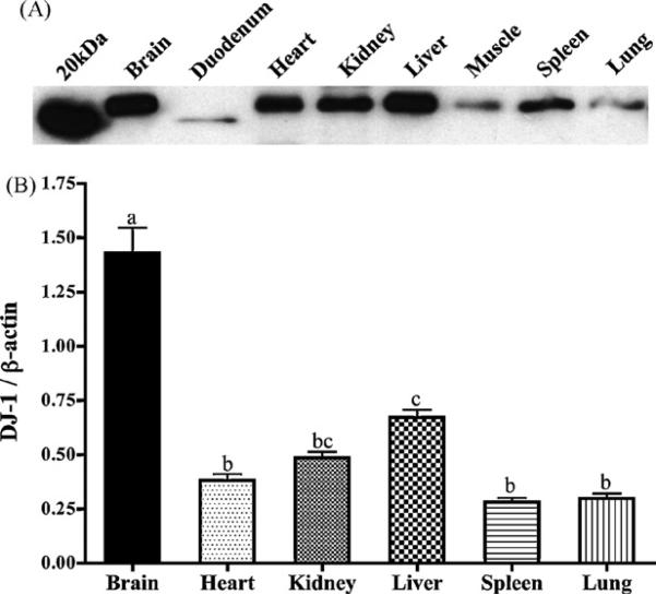
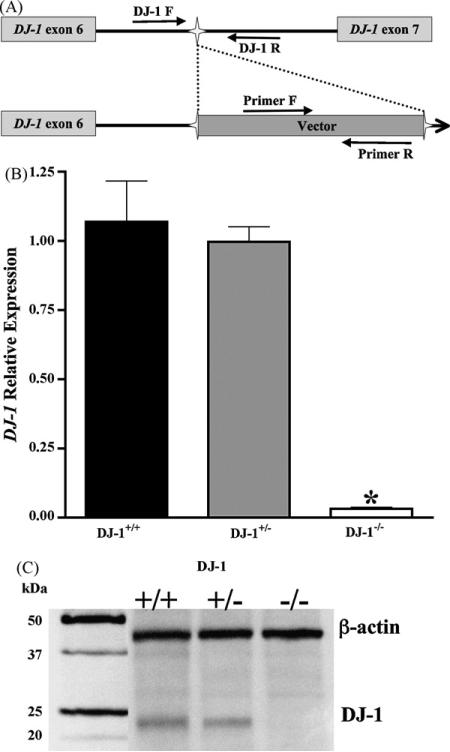
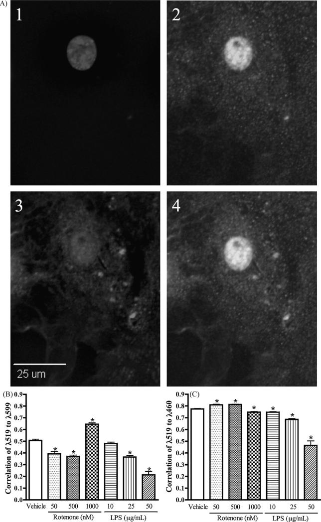
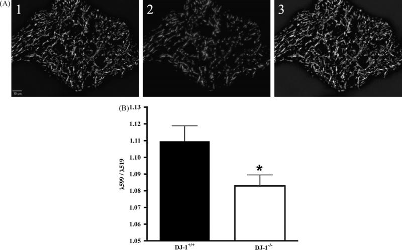
DJ-1 mutation induces early-onset Parkinson's disease, and conversely over-expression of DJ-1 is associated with cancer in numerous tissues. A gene-trap screening library conducted in embryonic stem cells was utilized for generation of a DJ-1 mutant mouse. Real-time PCR and immunoblotting were utilized to confirm functional mutation of the DJ-1 gene. Normal DJ-1 protein expression in adult mouse tissue was characterized and demonstrates high expression in brain tissue with wide systemic distribution. Primary astrocytes isolated from DJ-1(-/-) mice reveal a decreased nuclear localization of DJ-1 protein in response to rotenone or LPS, with a concomitant increase in mitochondrial localization of DJ-1 found only in the rotenone exposure. Resting mitochondrial membrane potential was significantly lower in DJ-1(-/-) astrocytes, as compared to controls. Our DJ-1 knockout mouse provides an exciting tool for exploring the molecular and physiological roles of DJ-1 to further explicate its functions in neurodegeneration.


Figures

Fig. 1
Fig. 1
DJ-1 protein is widely distributed in the mouse. (A) A representative blot depicts relative expression of DJ-1 in 25 μg of protein from each respective tissue. (B) DJ-1 protein levels were assayed and are expressed relative to β-actin in each tissue (n ≥ 4), and demonstrate high expression in brain tissue. Bars with differing letters are significantly different from one another (p < 0.05).
Fig. 2
Fig. 2
Gene-trap insertion into DJ-1 induces a functional mutation. (A) Schematic representation of the gene-trap in the endogenous DJ-1 gene. Following identification of the gene-trap insertion site, primers encompassing this sequence were designed, and, in combination with primers in the exogenous DNA, differentiation of DJ-1+/+, DJ-1+/−, or DJ-1−/− animals was accomplished. (B) DJ-1 mRNA expression was measured via real-time PCR in whole brain tissue. DJ-1−/− brain tissue has significantly decreased expression of DJ-1 compared to DJ-1+/+ or DJ-1+/− (n ≥ 4, p < 0.001). (C) DJ-1 protein expression is evident in DJ-1+/+ and DJ-1+/− animals, but absent in DJ-1−/−.
Fig. 3
Fig. 3
Rotenone increases mitochondrial localization of DJ-1, and both rotenone and LPS decrease nuclear localization. Primary cortical astrocytes were exposed to 50, 500 or 1000 nM rotenone + PBS, 10, 25, or 50 μg/mL LPS + DMSO, or control (PBS + DMSO) for 24 h, then loaded with MitoTracker Red and probed for DJ-1 immunofluorescence. To allow nuclear visualization, cells were mounted in DAPI-containing media. Pearson's correlation coefficients were calculated using SlideBook software image (A) A representative image used for analysis: (1) DAPI-stained nucleus, (2) DJ-1 immunofluorescence, (3) MitoTracker, and (4) merged image, for color image please see Supplementary Fig. 1. (B) DJ-1 correlation with the mitochondria (λ519:λ599) increased upon exposure to rotenone but not LPS (n ≥ 71, p < 0.001). (C) Nuclear correlation DJ-1 (λ519:λ460) decreased following exposure to either rotenone or LPS (n ≥ 71, p < 0.001).
Fig. 4
Fig. 4
Mutation of DJ-1 decreases resting mitochondrial membrane potential. Astrocytes were loaded with the ratiometric mitochondrial dye JC-1 (5 μM) for 30 min, then rinsed once in MEM with 10 mM HEPES without phenol red, and images were collected every 2 min for 8 min. Mitochondrial membrane potential, expressed as the ratio of λ599:λ519 was measured using SlideBook image analysis software. (A) Representative image of the mitochondrial dye JC-1, (1) fluorescence emitted at λ519, (2) fluorescence emitted at λ599, and (3) merged images of 1 and 2, for color image please see Supplementary Fig. 2. (B) DJ-1−/− astrocytes have decreased basal membrane potential compared to DJ-1+/+ (n = 48, p = 0.02).

Similar articles

Cited by

References

    1. Allen JW, Mutkus LA, Aschner M. Isolation of neonatal rat cortical astrocytes for primary cultures. Curr. Protoc. Toxicol. Suppl. 2000;4:12.14.11–12.14.15. - PubMed
    1. Aschner M, Gannon M, Kimelberg HK. Manganese uptake and efflux in cultured rat astrocytes. J. Neurochem. 1992;58:730–735. - PubMed
    1. Betarbet R, Canet-Aviles RM, Sherer TB, Mastroberardino PG, McLendon C, Kim JH, Lund S, Na HM, Taylor G, Bence NF, Kopito R, Seo BB, Yagi T, Yagi A, Klinefelter G, Cookson MR, Greenamyre JT. Intersecting pathways to neurodegeneration in Parkinson's disease: effects of the pesticide rotenone on DJ-1, alpha-synuclein, and the ubiquitin-proteasome system. Neurobiol. Dis. 2006;22:404–420. - PubMed
    1. Betarbet R, Sherer TB, MacKenzie G, Garcia-Osuna M, Panov AV, Greenamyre JT. Chronic systemic pesticide exposure reproduces features of Parkinson's disease. Nat. Neurosci. 2000;3:1301–1306. - PubMed
    1. Boka G, Anglade P, Wallach D, Javoy-Agid F, Agid Y, Hirsch EC. Immunocytochemical analysis of tumor necrosis factor and its receptors in Parkinson's disease. Neurosci. Lett. 1994;172:151–154. - PubMed

Publication types

MeSH terms