. 2009 Feb;40(2):280-92.
doi: 10.1016/j.mcn.2008.11.001. Epub 2008 Nov 19.

Neuronal Ca2+ sensor VILIP-1 leads to the upregulation of functional alpha4beta2 nicotinic acetylcholine receptors in hippocampal neurons

Affiliations

Neuronal Ca2+ sensor VILIP-1 leads to the upregulation of functional alpha4beta2 nicotinic acetylcholine receptors in hippocampal neurons

C J Zhao et al. Mol Cell Neurosci. 2009 Feb.

Abstract

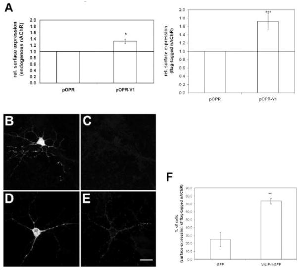
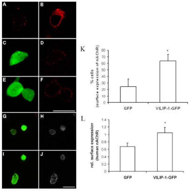
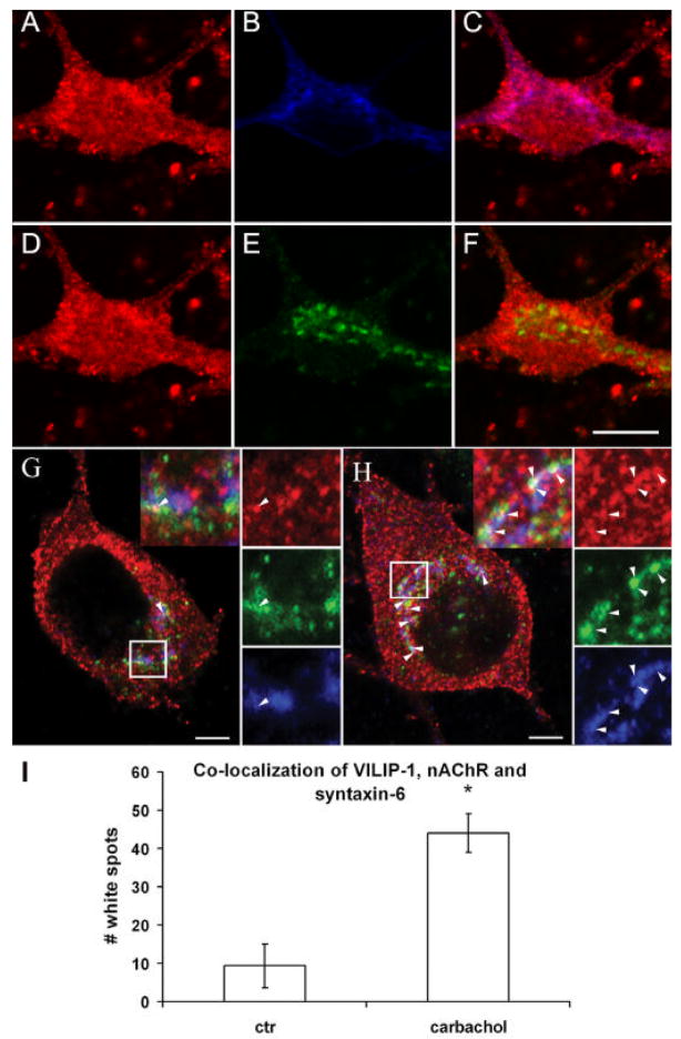
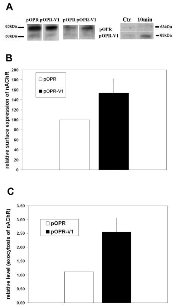
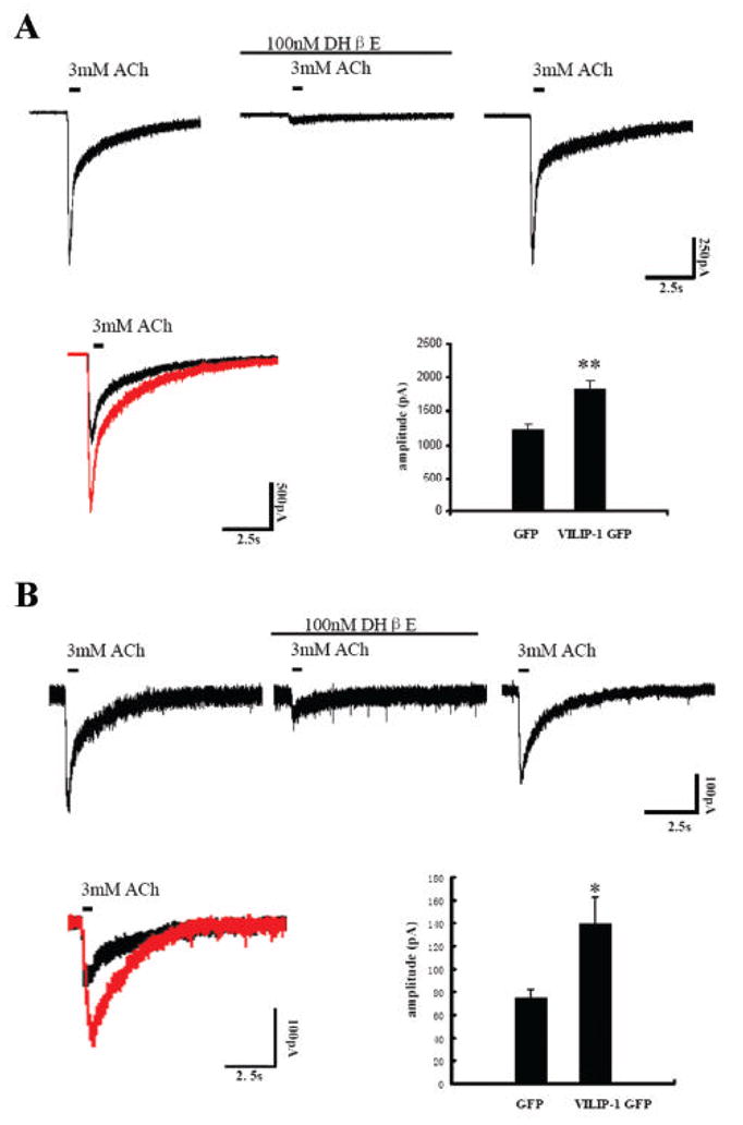
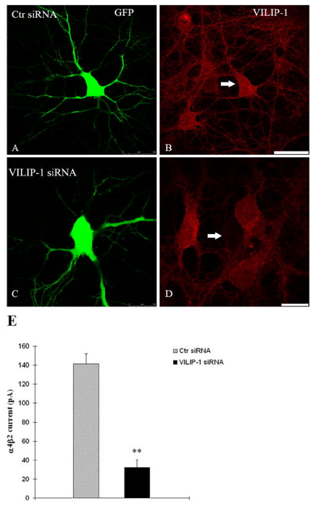
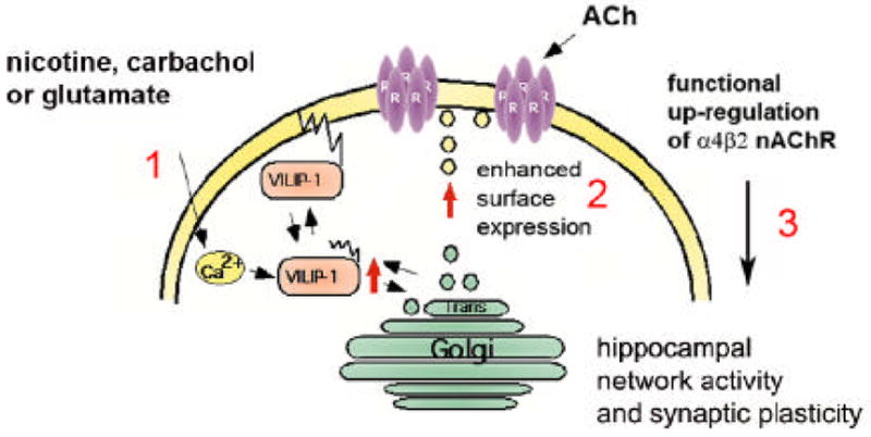
The neuronal Ca2+-sensor protein VILIP-1, known to affect clathrin-dependent receptor trafficking, has been shown to interact with the cytoplasmic loop of the alpha4-subunit of the alpha4beta2 nicotinic acetylcholine receptor (nAChR), which is the most abundant nAChR subtype with high-affinity for nicotine in the brain. The alpha4beta2 nAChR is crucial for nicotine addiction and the beneficial effects of nicotine on cognition. Its dysfunction has been implicated in frontal lobe epilepsy, Alzheimer's disease and schizophrenia. Here we report that overexpression of VILIP-1 enhances ACh responsiveness, whereas siRNA against VILIP-1 reduces alpha4beta2 nAChR currents of hippocampal neurons. The underlying molecular mechanism likely involves enhanced constitutive exocytosis of alpha4beta2 nAChRs mediated by VILIP-1. The two interaction partners co-localize in a Ca2+-dependent manner with syntaxin-6, a Golgi-SNARE protein involved in trans-Golgi membrane trafficking. Thus, we speculate that regulation of VILIP-1-expression might modulate surface expression of ligand-gated ion channels, such as the alpha4beta2 nAChRs, possibly comprising a novel form of physiological up-regulation of ligand-gated ion channels.


Figures

Fig. 1
Fig. 1. Expression of VILIP-1 enhances surface expression of endogenous and co-transfected α4β2 nACh receptor in hippocampal neurons
A. VILIP-1 cDNA (pOPR-V1) or control vector (pOPR) was transfected alone (left) or co-transfected with flag-tagged α4β2 nAChR (right) into two week old primary hippocampal cultures. 16h following transfection, where no changes in cell numbers were observed, the surface expression of the endogenous α4β2 nAChR (left) or of the flag-tagged α4β2 nAChR (right) was quantified using mab299 against the α4-subunit (left) or an anti-flag antibody (right) in ELISA. BE. Control GFP vector (B, C) or VILIP-1-GFP cDNA (D, E) were co-transfected with flag-tagged α4β2 nAChR (B–E) into 2 week old primary hippocampal cultures. After 40h in culture surface expression of the flag-tagged α4β2 nAChR (C, E) was monitored with anti-flag antibody by confocal immunofluorescence microscopy. Bar in E is 20 μm. F. Quantification of the number of cells expressing flag-tagged α4β2 nAChR at the cell surface in GFP (C) and VILIP-1-GFP (E) co-transfected hippocampal neurons. Mean values ± S.D. are from five experiments for A and three experiment for F carried out in triplicate.
Fig. 2
Fig. 2. Expression of VILIP-1 enhances surface expression of transiently transfected α4β2 nAChR in HEK cells and of human α4β2 nAChR in stably transfected HEK cells. A–F
Flag-tagged α4β2 nAChR was transfected alone (A, B) or co-transfected with GFP (C, D), or with VILIP-1-GFP cDNA (E, F) into HEK cells. After 48h in culture only little surface expression of the flag-tagged α4β2 nAChR was monitored (A), but the receptor was strongly expressed intracellularly when stained in the presence of TX100 (B). Surface expression in GFP (C, D) and VILIP-1-GFP (E, F) co-transfected HEK cells was monitored with anti-flag antibody by confocal immunofluorescence microscopy (D, F). Bar in F is 20 μm. GJ. GFP (G, H) or VILIP-1-GFP cDNA (I, J) was transfected into HEK cells stably expressing the human α4β2 nAChR. After 48h in culture surface expression of human α4β2 nAChR (H, J) was monitored by confocal immunofluorescence microscopy in GFP (G, H) and VILIP-1-GFP (I, J) transfected cells with mAb299 antibody against the α-subunit of nAChR (H, J). Cross-bleeding of channels was avoided by sequential analysis of the red and green channel. Bar in J is 20 μm. K. Quantification of the number of cells expressing flag-tagged α4β2 nAChR in GFP (D) and VILIP-1-GFP (F) co-transfected HEK cells. No differences in cell numbers between GFP- and GFP-VILIP-1-transfected cells was observed. L. Quantification of the surface expressing of human α4β2 nAChR in GFP (H) and VILIP-1-GFP (J) transfected HEK cells in mAb299 stained cultures using the NIH ImageJ program. Mean values ± S.D. are from three experiments carried out in triplicate.
Fig. 3
Fig. 3. Ca2+-dependent localization of VILIP-1 to cell surface and the intracellular membranes of Golgi and ER in hippocampal neurons
AL. Hippocampal neurons were unstimulated (A–C, G–I) or stimulated with carbachol (D–F, J–L), which increases intracellular Ca2+ concentrations to enhance the membrane association of VILIP-1 (compare A to D and G to J), as described in detail previously for ionomycin treatment (28) AF. Co-localization with the ER marker calnexin (B, E) could not be found in the merged images (C, F, VILIP-1: red, calnexin: green). GL. Co-staining with the trans-Golgi marker syntaxin-6 (H, K) was observed in yellow in the merged images (I, L), and was enhanced after carbachol treatment to increase intracellular calcium (L), as previously described in detail for ionomycin treatment (Spilker et al., 2002). Representative stainings out of at least three experiments carried out in triplicate are shown. Bar in M is 20 μm.
Fig. 4
Fig. 4. Co-localization of α4β2 nAChR, ER and Golgi marker in a triple co-stained hippocampal neuron
A–F. Hippocampal neurons cultured for two weeks transfected with flag-tagged α4β2 nAChR were simultaneously stained with anti-flag antibody and antibodies against syntaxin-6 and calnexin. A–C. Co-localization of α4β2 nAChR (A) with the ER marker calnexin (B) is seen in purple in the merged picture (C). D–F. Occasional co-localization of α4β2 nAChR (D) with the Golgi marker syntaxin-6 (E) is seen in yellow in the merged picture (F). Note, that no co-localization of Golgi marker syntaxin-6 can be found with α4β2 nAChR and the ER marker calnexin which would be observed as white in merged pictures (data not shown). Bar in F is 20 μm. Ca2+-dependent co-localization of VILIP-1-GFP, α4β2 nAChR and the Golgi marker syntaxin-6 in a triple co-stained hippocampal neuron. G, H. Hippocampal neurons cultured for two weeks were transfected with flag-tagged α4β2 nAChR and VILIP-1-GFP and simultaneously stained with anti-flag antibody and antibodies against syntaxin-6. G. Co-localization of VILIP-1-GFPα4β2 nAChR with the Golgi marker syntaxin-6 is seen occasionally (arrow) without stimulation as white co-staining in the merged picture (G, upper inset: VILIP-1, middle inset: α4β2 nAChR, lower inset: syntaxin-6, white box shows the magnified area above). H. In carbachol treated cultures, substantial co-localization of all three markers is observed as white co-staining in distinct spots within the hippocampal neuron (H, arrows point at white co-localization spots, white box shows the magnified area with highest density of co-localization). Bar in A, B is 5 μm. I. Quantification of the co-localization of VILIP- 1-GFP, α4β2 nAChR and syntaxin-6 as number of white spots in control (G) carbachol-treated (H) triple co-stainings. Mean values ± S.D. are from three experiments carried out in duplicate.
Fig. 5
Fig. 5. Expression of VILIP-1 affects surface expression via accelerating exocytosis of the α4β2 nAChR in double-transfected hippocampal neurons and in HEK cells
A. In HEK cells co-transfected with flag-tagged α4β2 nAChR and pOPR-VILIP-1 or pOPR vector alone, biotinylation of cellular proteins and of surface proteins was performed and the flag-tagged α4β2 nAChR was detected in Western blot analysis of streptavidin-precipitates from the cell extracts. For input controls (left panel) following lysis and biotinylation, or for surface protein determination (middle panel) following biotinylation and then lysis, the flag-immunoreactive bands in streptavidin-precipitates at about 70 and 55kDa representing α4 and β2 subunits are monitored by Western blot analysis of control vector (pOPR) and VILIP-1-transfected (pOPR-V1) HEK cell extracts. Exocytosis was determined after enzymatic digest of surface proteins and subsequent biotinylation and lysis. The re-appearance of the α4–subunit signal in the streptavidin-precipitates was measured by biotinylation 10min after enzymatic treatment (right panel). B, C. Quantification of the Western blot in A, middle panel, indicates (B) a significant 50% increase in relative surface expression and (C) a 2,5 fold increase in relative level of exocytosis, in A, right panel, of the α4–subunit of the nAChR in VILIP-1-transfected HEK cells.
Fig. 6
Fig. 6. Potentiation of ACh-induced whole-cell currents by expression of VILIP-1-GFP in HEK293 cells stably expressing human α4β2 nAChR (A) and in hippocampal neurons transfected with rat α4β2 nAChR (B)
A. Whole-cell responses to ACh in the absence and presence of 100nM DHβE recorded from a single cell of the Ha4b2Lx/1 cell line, which was held at -60mV. The ACh concentration applied (3mM) was in the range of the saturation response. ACh evoked current was blocked completely by 100nM of the α4β2 nAChR antagonist DHβE (second trace). The current recovered after DHβE wash out. Representative traces of whole-cell responses following ACh stimulation in GFP (black trace) and GFP-VILIP-1 (red trace) transfected HEK cells are shown in the lower left panel. The response to ACh in VILIP-1-GFP transfected cells (lower right panel, n=33, from two experiments) is highly significant increased in amplitude (p<0.01) compared to the responses to ACh in GFP-transfected cells (n=18, from two experiments). B. Whole-cell responses to ACh, in the absence and presence of 100nM DHβE recorded from a single cultured hippocampal neuron which was held at -60mV. The evoked current partially recovered after DHβE wash out. Representative traces of whole-cell responses following ACh stimulation in GFP (black trace) and GFP-VILIP-1 (red trace) transfected neurons are shown in the left panel. The response to ACh in the VILIP-1-GFP transfected cells (lower right panel, n=13, from two experiments) shows highly significant increase in amplitude (p<0.05), as compared to GFP-transfected neurons (n=14, from three experiments).
Fig 7
Fig 7. SiRNA knockdown of VILIP-1 in GFP co-transfected hippocampal neurons (A–D) leads to reduction of ACh-induced whole-cell α4β2 nAChR currents
Representative confocal image showing control siRNA and GFP co-transfected hippocampal neuron (A) and expression of endogenous VILIP-1 (B). VILIP-1-siRNA/GFP-coexpressing neurons (C) show strongly reduced expression of endogenous VILIP-1 (D). In the representative experiment shown an average of 13 out of 15 co-transfected neurons per dish showed strongly reduced VILIP-1 expression levels, whereas 29 out of 33 neurons in the control siRNA group did not show changes in expression. Bar in A, B is 30μm, bar in C,D is 15 μm. E. In hippocampal neurons co-transfected with α4β2 nAChRs and control siRNA (Ctr siRNA) the average α4 nicotinic current amplitude was strongly reduced following co-transfection with VILIP-1 siRNA (VILIP-1 siRNA). Data are presented as mean ± SEM; ** p < 0.01 compared with control group.
Fig. 8
Fig. 8. Hypothesis for a novel physiological mechanism of up-regulation of functional α4β2
The cartoon shows the neuronal Ca2+-sensor VILIP-1, is activated following stimulation of a neuron by a signal, such as glutamate, carbachol or nicotine, which is acting on receptors increasing the intracellular Ca2+ level (1). VILIP-1 shuttles to cell surface and Golgi membranes as well as clathrin-coated vesicles, where it co-localizes with α4β2 nAChR and with syntaxin-6, a SNARE implicated in clathrin-dependent transport mechanisms in the trans-Golgi network. VILIP-1 causes enhanced exocytosis and surface transport of α4β2 nAChR (2), consequently increasing the surface expression and finally the sensitivity of the neuron towards ACh (3). The nicotine or glutamate induced translocation of VILIP-1 to cellular membranes (1, Zhao et al., 2008), and in turn the up-regulation of functional α4β2 nAChRs (2, Lin et al., 2002, this study) may contribute to plasticity of nicotinergic neurotransmission in principal neurons and/or in interneurons in the hippocampus, since the expression of VILIP-1 in conjunction with the α4β2 nAChR enhances the frequency of inhibitory postsynaptic currents (IPSCs) in hippocampal cultures (3, Gierke et al., 2008). Thereby, the physiological up-regulation of α4β2 nAChR by VILIP-1 might modulate hippocampal network activity and synaptic plasticity.

References

    1. Ames JB, Ishima R, Tanaka T, Gordon JI, Stryer L, Ikura M. Molecular mechanics of calcium-myristoyl switches. Nature. 1997;389:198–202. - PubMed
    1. Bernstein HG, Becker A, Keilhoff G, Gorcyca WA, Braunewell KH, Grecksch G. Ketamine induced changes of the expression of neuronal calcium sensor proteins VILIP-1, -3 and hippocalcin in a partial schizophrenia model in rats. Neurosci Lett. 2003;339:95–98. - PubMed
    1. Bernstein HG, Braunewell KH, Spilker C, Danos P, Baumann B, Diekmann S, Gundelfinger ED, Bogerts B. Hippocampal expression of the Ca2+ sensor protein VILIP-1 in schizophrenia. Neuroreport. 2002;23:393–396. - PubMed
    1. Blondeau F, Ritter B, Allaire PD, Wasiak S, Girard M, Hussain NK, Angers A, Legendre-Guillemin V, Roy L, Boismenu D, Kearney RE, Bell AW, Bergeron JJ, McPherson PS. Tandem MS analysis of brain clathrin-coated vesicles reveals their critical involvement in synaptic vesicle recycling. Proc Natl Acad Sci U S A. 2004;101:3833–3838. - PMC - PubMed
    1. Bock JB, Klumperman J, Davanger S, Scheller RH. Syntaxin 6 functions in trans- Golgi network vesicle trafficking. Mol Biol Cell. 1997;8:1261–1271. - PMC - PubMed

Publication types

MeSH terms