. 2009 Apr 12;364(1519):991-1003.
doi: 10.1098/rstb.2008.0260.

Zebrafish and medaka: model organisms for a comparative developmental approach of brain asymmetry

Affiliations

Zebrafish and medaka: model organisms for a comparative developmental approach of brain asymmetry

Iskra A Signore et al. Philos Trans R Soc Lond B Biol Sci. .

Abstract

Comparison between related species is a successful approach to uncover conserved and divergent principles of development. Here, we studied the pattern of epithalamic asymmetry in zebrafish (Danio rerio) and medaka (Oryzias latipes), two related teleost species with 115-200 Myr of independent evolution. We found that these species share a strikingly conserved overall pattern of asymmetry in the parapineal-habenular-interpeduncular system. Nodal signalling exhibits comparable spatial and temporal asymmetric expressions in the presumptive epithalamus preceding the development of morphological asymmetries. Neuroanatomical asymmetries consist of left-sided asymmetric positioning and connectivity of the parapineal organ, enlargement of neuropil in the left habenula compared with the right habenula and segregation of left-right habenular efferents along the dorsoventral axis of the interpeduncular nucleus. Despite the overall conservation of asymmetry, we observed heterotopic changes in the topology of parapineal efferent connectivity, heterochronic shifts in the timing of developmental events underlying the establishment of asymmetry and divergent degrees of canalization of embryo laterality. We offer new tools for developmental time comparison among species and propose, for each of these transformations, novel hypotheses of ontogenic mechanisms that explain interspecies variations that can be tested experimentally. Together, these findings highlight the usefulness of zebrafish and medaka as comparative models to study the developmental mechanisms of epithalamic asymmetry in vertebrates.


Figures
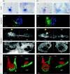
Figure 1
Figure 1
(a) Zebrafish and (b) medaka share an overall pattern of molecular and morphological epithalamic asymmetry. ((i),(ii)) Asymmetric expression of components of the nodal signalling pathway in the presumptive epithalamus. mRNA expression of orthologue genes was detected by whole-mount in situ hybridization (arrows) at (i) normalized STU 35 (Dr-lefty1, Ol-lefty) and (ii) 43 (Dr-pitx2c, Ol-pitx2c) (table 2). The lateral flexure of the third ventricle is indicated by arrowheads. (iii) Left-sided positioning and efferent projection of the parapineal organ. Expression of GFP was detected in medaka Tg(fRx2::GFP) and zebrafish Tg(FoxD3::GFP) and pseudo-coloured in blue and green to label pineal and parapineal organs, respectively. The red arrowhead points to the terminal dandelion seed-head-shaped domain of parapineal efferent connectivity. (iv) Asymmetric organization of neuropil in the habenulae. Arrows indicate the regions of neuropil, which exhibit dissimilar growth between the left and right habenulae, as detected by immunostaining against acetylated α-tubulin. The red arrowhead points to a neuropil domain that is detected exclusively in the left habenula of medaka. (v) Asymmetric organization of neuronal cell bodies in the habenulae. Asterisks indicate equivalent regions of the left and right habenulae. The red arrowhead points to a domain devoid of fluorescent Nissl staining that is detected exclusively in the left habenula of medaka. ((vi),(vii)) Dorsoventral segregation of left–right habenular efferents in the IPN. Left and right habenular projections were labelled with DiD (red) and DiO (green), respectively. Images correspond to ((i)–(vi)) dorsal and (vii) lateral views with anterior and dorsal to the top, respectively. Stages of development correspond to 120 HPF (zebrafish, at 26°C) and Iwamatsu's stage 39 (medaka), unless otherwise stated. ((iii)–(vii)) Three-dimensional projections from confocal z-stacks. L, left; R, right; Lh, left habenula; Rh, right habenula; hc, habenular commissure; Lfr, left fasciculus retroflexus; Rfr, right fasciculus retroflexus; dIPN, dorsal domain of the IPN; vIPN, ventral domain of the IPN. Scale bars, ((i)–(v)) 20 μm, ((vi),(vii)) 30 μm.
Figure 2
Figure 2
Heterotopic parapineal efferent connectivity in the left habenula of (a) zebrafish and (b) medaka. (a(i)–(vi)) Parapineal efferents blend into a dorsomedial neuropil domain of the left habenula in zebrafish whereas in (b(i)–(vi)) they segregate from other sources of habenular neuropil to form a distinct dorso-anteromedial domain in the left habenula of medaka. Images correspond to dorsal views of the left habenula with anterior to the top. Images were obtained after three-dimensional maximum projections from confocal z-stacks. The parapineal organ was pseudo-coloured in blue (parapineal body) and green (parapineal efferents) after immunostaining against GFP in (a(i),(iv)) 120 HPF zebrafish Tg(FoxD3::GFP) and (b(i),(iv)) St.39 medaka Tg(fRx2::GFP). Distribution of neuropil and nuclei in the left habenula were detected by (ii) immunostaining against acetylated α-tubulin and (v) fluorescent To-pro staining, respectively. Merged images of double labelling are shown in the bottom panels ((iii),(vi)). Asterisks indicate nuclei-free domains of the left habenula where parapineal connectivity is distributed in zebrafish. Arrowheads point to the terminal dandelion seed-head-shaped domain of parapineal efferent connectivity in medaka. (vii) Summary model of parapineal efferent connectivity in zebrafish and medaka. (a(vii)) In zebrafish, parapineal efferents distribute broadly within a large dorsomedial neuropil domain of the left habenula situated immediately anterior to the habenular commissure. (b(vii)) In medaka, parapineal efferents form a thick bundle of axons, which after entering the left habenula, make a turn towards the midline to end in a well-defined dandelion seed-head-shaped neuropil domain situated in the most dorso-anteromedial aspect of the left habenula. All images correspond to dorsal views, with anterior to the top. The body of the parapineal organ and its efferent connectivity are shown in black, the habenular commissure in grey and neuropil domains in yellow. L, left; R, right; Lh, left habenula; Rh, right habenula; hc, habenular commissure. Scale bars, 20 μm.
Figure 3
Figure 3
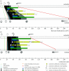
Comparison of sequence, relative timing and duration of developmental events during the establishment of epithalamic asymmetry in zebrafish and medaka. The diagrams show the temporal occurrence of key steps of asymmetric brain morphogenesis in zebrafish and medaka, expressed in (a) absolute and (b) normalized times. To provide a contextual view, the timing of main embryonic events is also included. The colour codes shown at the bottom of the figure indicate different developmental events (lines) and periods (boxes or bars) analysed in the temporal plots of (a,b). For clarity, equivalent events in medaka and zebrafish are joined. Diagrams of developmental stages were obtained from the literature (Kimmel et al. 1995; Iwamatsu 2004). Schematic of epithalamic asymmetry events (bottom right) correspond, from top to bottom, to: a frontal view of the epithalamus depicting left-sided asymmetric nodal expression, a dorsal view of the pineal complex showing the initiation of left-sided parapineal axonal projection and a dorsal view of the IPN (white circle) revealing habenular efferent connectivity reaching dorsal and ventral regions of the IPN. The scale was maintained in (a,b) to emphasize the effect of time normalization. Zebrafish and medaka show a conserved sequence of developmental events of epithalamic asymmetry although they exhibit distinct relative timing (heterochrony).
Figure 4
Figure 4
Developmental models of heterotopic parapineal efferent connectivity. (a) Zebrafish: asymmetries of the parapineal organ and habenulae interact in three consecutive steps during development. (i) Initial left–right biases in the presumptive habenular region guide parapineal migration to the left side. (ii) Subsequent left-sided positioning of the parapineal organ is involved in the induction/propagation of a distinct spatial pattern of habenular differentiation. (iii) Finally, the topology of subdomains arising after habenular differentiation determines the position of habenular neurons receiving parapineal efferent connectivity. (b) Medaka (model 1): heterotopic parapineal efferent connectivity arises from a different topological organization of habenular subdomains. In this model, spatial differences in the location of the parapineal organ at the time of habenular differentiation and/or underlying differences in habenular (i) pre-patterning lead to (ii) distinct topological programmes of habenular subdomain differentiation and (iii) subsequent positioning of parapineal target cells. (c) Medaka (model 2): heterotopic parapineal efferent connectivity arises from different selection of parapineal target cells within the habenulae. In this model, parapineal migration and habenular differentiation are equivalent in both species. However, parapineal projections reach different target neurons in the habenula of both species owing to the differences in axon guidance cues. (i) Shaded green regions depict molecular left–right biases within the presumptive habenulae. The movement of the parapineal organ from the midline to the left side (arrows) is represented as partially overlapping drawings of parapineal outlines. (ii) White and red regions illustrate putative subdomains of the habenulae. Arrows illustrate the direction of the inductive properties of the parapineal organ. (iii) The topological pattern of parapineal efferent connectivity (black) and the location of parapineal target cells within the habenula (colours) are shown (see also figure 2). Colours represent equivalent cellular identities. All diagrams correspond to dorsal views, with anterior to the top. For clarity, only the left habenula is illustrated, and the right habenula is depicted with dotted lines. L, left; R, right; Lh, left habenula; Rh, right habenula; hc, habenular commissure.

References

    1. Aizawa H., Bianco I.H., Hamaoka T., Miyashita T., Uemura O., Concha M.L., Russell C., Wilson S.W., Okamoto H. Laterotopic representation of left–right information onto the dorso-ventral axis of a zebrafish midbrain target nucleus. Curr. Biol. 2005;15:238–243. doi:10.1016/j.cub.2005.01.014 - DOI - PMC - PubMed
    1. Aizawa H., Goto M., Sato T., Okamoto H. Temporally regulated asymmetric neurogenesis causes left–right difference in the zebrafish habenular structures. Dev. Cell. 2007;12:87–98. doi:10.1016/j.devcel.2006.10.004 - DOI - PubMed
    1. Barth K.A., Miklosi A., Watkins J., Bianco I.H., Wilson S.W., Andrew R.J. fsi zebrafish show concordant reversal of laterality of viscera, neuroanatomy, and a subset of behavioral responses. Curr. Biol. 2005;15:844–850. doi:10.1016/j.cub.2005.03.047 - DOI - PMC - PubMed
    1. Bianco I.H., Wilson S.W. The habenular nuclei: a conserved asymmetric relay station in the vertebrate brain. Phil. Trans. R. Soc. B. 2009;364:1005–1020. doi:10.1098/rstb.2008.0213 - DOI - PMC - PubMed
    1. Bianco I.H., Carl M., Russell C., Clarke J.D., Wilson S.W. Brain asymmetry is encoded at the level of axon terminal morphology. Neural Dev. 2008;3:9. - PMC - PubMed

Publication types

MeSH terms

LinkOut - more resources