Review
. 2009 Apr 12;364(1519):1005-20.
doi: 10.1098/rstb.2008.0213.

The habenular nuclei: a conserved asymmetric relay station in the vertebrate brain

Affiliations
Review

The habenular nuclei: a conserved asymmetric relay station in the vertebrate brain

Isaac H Bianco et al. Philos Trans R Soc Lond B Biol Sci. .

Abstract

The dorsal diencephalon, or epithalamus, contains the bilaterally paired habenular nuclei and the pineal complex. The habenulae form part of the dorsal diencephalic conduction (DDC) system, a highly conserved pathway found in all vertebrates. In this review, we shall describe the neuroanatomy of the DDC, consider its physiology and behavioural involvement, and discuss examples of neural asymmetries within both habenular circuitry and the pineal complex. We will discuss studies in zebrafish, which have examined the organization and development of this circuit, uncovered how asymmetry is represented at the level of individual neurons and determined how such left-right differences arise during development.


Figures

Figure 1
Figure 1
Connectivity of the DDC. This schematic shows the principal connections of the medial and lateral habenulae and interpeduncular nucleus as described in mammals, in particular the rat. Thick arrows highlight the septum–MHb–IPN axis and the convergence of limbic and striatal inputs into the lateral habenula. Notably, there are very limited data regarding the relative functional importance of the various connections shown here. Acb, nucleus accumbens; Circad., potential sources of circadian information; CPu, caudate/putamen; Dors. Teg. region, dorsal tegmental region; DBB, nucleus of diagonal band; DTN, ventral tegmental nucleus of Güdden; EP, entopeduncular nucleus; FrCx, frontal cortex; HippoF, hippocampal formation; IPN, interpeduncular nucleus; LC, locus coeruleus; LH, lateral hypothalamic area; LHb, lateral habenula; LPO, lateral preoptic area; MHb, medial habenula; NI, nucleus incerta; P, pineal; SC, superior colliculus; SCN, suprachiasmatic nucleus; SNc, substantia nigra pars compacta; sf, septofimbrial nucleus; Thal, thalamic nuclei; tr, nucleus triangularis; VTA, ventral tegmental area; VTN, ventral tegmental nucleus of Güdden.
Figure 2
Figure 2
Asymmetric habenular circuitry in zebrafish. (a) Schematic showing connectivity of the habenular complex in larval zebrafish. A significant afferent input derives from migrated neurons of the eminentia thalami, which is thought to form the entopeduncular/peripeduncular complex in adult zebrafish (note that the teleostean entopeduncular complex is not part of the pallidum and does not correspond to the EP of amniotes; Wullimann & Mueller 2004). EmT neurons project bilaterally, innervating both left and right habenulae. A subset of left- and right-sided neurons in the anterior pallium are a source of asymmetric innervation, selectively terminating in a small medial domain of the right habenula (indicated in purple). In addition, a small afferent input may derive from the posterior tuberculum (Hendricks & Jesuthasan 2007). In the epithalamus, the left-sided parapineal exclusively innervates the left habenula (Concha et al. 2003). Habenular neurons project efferent axons that course in the fasciculus retroflexus. A major target is the interpeduncular nucleus: left- and right-sided axons are segregated along the dorso-ventral (DV) axis of the IPN in a laterotopic manner (Aizawa et al. 2005). A smaller and apparently symmetric contingent of habenular axons terminates caudal to the IPN in the serotonergic raphe. (b) Neuroanatomical asymmetries in the dorsal diencephalon. Anti-acetylated tubulin immunostaining (red) shows that the left habenula contains a greater density of neuropil, especially in the dorsomedial aspect of the nucleus. The pineal (blue) and parapineal (green) are visualized by the expression of green fluorescent protein (GFP) in a Tg(foxD3:GFP) transgenic larva. The parapineal is asymmetric in both its location and connectivity, and its efferent axons preferentially terminate in the asymmetric medial neuropil of the left habenula. Dorsal view, anterior top. (c) Three-dimensional confocal reconstruction showing habenular axon terminals in the ventral midbrain labelled using lipophilic tracer dyes applied to the habenulae. Left-sided axons were labelled with DiD (red) and right-sided axons with DiI (green). The dorsal IPN is almost exclusively innervated by left-sided axons, whereas the ventral target receives a majority of right-sided inputs. Dorsal view, anterior top. Tel, telencephalon; EmT, eminentia thalami; PT, posterior tuberculum; Pa, pallium; sm, stria medullaris; Hb, habenula; hc, habenular commissure; pp, parapineal, P, pineal; pc, posterior commissure; FR, fasciculus retroflexus; TeO, optic tectum; IPN, interpeduncular nucleus; a, anterior; p, posterior; l, left; r, right; d, dorsal; v, ventral. Adapted from Bianco et al. (2008). A number of these asymmetry phenotypes are conserved in the distantly related teleost medaka (Oryzias latipes; Signore et al. 2009).
Figure 3
Figure 3
Zebrafish Hb–IPN projection neurons elaborate one of two distinct axon terminal arbour morphologies. (a) Three-dimensional reconstruction showing a single right habenular projection neuron that was labelled by focal electroporation with a construct driving the expression of membrane GFP, in an intact larval zebrafish brain (4 days post-fertilization). The cell body, located in the right habenula (rHb) extends an axon down the right FR (indicated by arrow) that terminates in the IPN. Habenular neurons elaborate remarkable axon arbours within the IPN that cross the ventral midline multiple times. Scale bar, 100 μm. (b(i),c(i)) Dorsal and (b(ii),c(ii)) lateral confocal reconstructions of single habenular axon arbours in the IPN. (b(i),(ii)) Example of an L-typical axon arbour, formed by 84% of left habenular neurons. These arbours are located in the dorsal IPN and are shaped similar to a domed crown and arborize over a considerable dorsoventral extent (compare dorsal (b(i)) and lateral (b(ii)) views of an example L-typical arbour). (c(i),(ii)) Example of an R-typical axon arbour, which is considerably flatter, localized to the ventral IPN and formed by 90% of right habenular neurons. Adapted from Bianco et al. (2008).
Figure 4
Figure 4
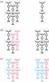
Models for lateralization of neural tissue. (a) Equivalent regions on the left and right of the CNS are identical in composition and differ only in overall size. (b) Unique types of neuron, or patterns of connectivity, may be specified on either the left or right or both sides (indicated by unique red neurons on the left in this schematic). (c) Identical circuit components might exist on both sides of the CNS, but in different ratios. Note that these models are in no way mutually exclusive. In fact, it is likely that all three strategies may be involved in the lateralization of DDC circuitry (see the main text).

References

    1. Adrio F., Anadon R., Rodriguez-Moldes I. Distribution of choline acetyltransferase (ChAT) immunoreactivity in the central nervous system of a chondrostean, the Siberian sturgeon (Acipenser baeri) J. Comp. Neurol. 2000;426:602–621. doi:10.1002/1096-9861(20001030)426:4<602::AID-CNE8>3.0.CO;2-7 - DOI - PubMed
    1. Aizawa H., Bianco I.H., Hamaoka T., Miyashita T., Uemura O., Concha M.L., Russell C., Wilson S.W., Okamoto H. Laterotopic representation of left–right information onto the dorso-ventral axis of a zebrafish midbrain target nucleus. Curr. Biol. 2005;15:238–243. doi:10.1016/j.cub.2005.01.014 - DOI - PMC - PubMed
    1. Aizawa H., Goto M., Sato T., Okamoto H. Temporally regulated asymmetric neurogenesis causes left–right difference in the zebrafish habenular structures. Dev. Cell. 2007;12:87–98. doi:10.1016/j.devcel.2006.10.004 - DOI - PubMed
    1. Andrew R.J., Osorio D., Budaev S. Light during embryonic development modulates patterns of lateralization strongly and similarly in both zebrafish and chick. Phil. Trans. R. Soc. B. 2009;364:983–989. doi:10.1098/rstb.2008.0241 - DOI - PMC - PubMed
    1. Araki M., McGeer P.L., Kimura H. The efferent projections of the rat lateral habenular nucleus revealed by the PHA-L anterograde tracing method. Brain Res. 1988;441:319–330. doi:10.1016/0006-8993(88)91410-2 - DOI - PubMed

Publication types

MeSH terms

LinkOut - more resources