. 2008 Dec 15;183(6):1075-88.
doi: 10.1083/jcb.200806035. Epub 2008 Dec 8.

Plasma membrane microdomains regulate turnover of transport proteins in yeast

Affiliations

Plasma membrane microdomains regulate turnover of transport proteins in yeast

Guido Grossmann et al. J Cell Biol. .

Abstract

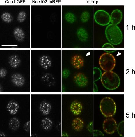
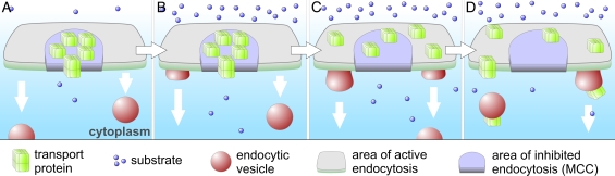
In this study, we investigate whether the stable segregation of proteins and lipids within the yeast plasma membrane serves a particular biological function. We show that 21 proteins cluster within or associate with the ergosterol-rich membrane compartment of Can1 (MCC). However, proteins of the endocytic machinery are excluded from MCC. In a screen, we identified 28 genes affecting MCC appearance and found that genes involved in lipid biosynthesis and vesicle transport are significantly overrepresented. Deletion of Pil1, a component of eisosomes, or of Nce102, an integral membrane protein of MCC, results in the dissipation of all MCC markers. These deletion mutants also show accelerated endocytosis of MCC-resident permeases Can1 and Fur4. Our data suggest that release from MCC makes these proteins accessible to the endocytic machinery. Addition of arginine to wild-type cells leads to a similar redistribution and increased turnover of Can1. Thus, MCC represents a protective area within the plasma membrane to control turnover of transport proteins.


Figures

Figure 1.
Figure 1.
10 new proteins sharing the MCC localization. Cortical distributions of 10 proteins (left; green in merge) were colocalized with the MCC pattern marked with Sur7-mRFP (middle; red in merge). Tangential confocal sections are presented showing the cell surface and fluorescence intensity profiles (diagrams) measured along the arrows. Mean filter was applied on the plotted curves to reduce the noise present in the raw data. Red and green curves were normalized to the same maximum value. Bar, 5 μm.
Figure 2.
Figure 2.
Distribution of MCC markers in selected knockout strains. Distributions of HUP1-GFP, Can1-GFP, Sur7-GFP, and filipin-stained sterols were monitored in the library of single gene deletion strains (see Materials and methods). Examples of detected phenotypes (classification of phenotypes: wild type [WT]–like, −; weak, +; medium, ++; strong, +++) on tangential confocal sections (HUP1, Can1, and Sur7) or wide-field images (filipin; transversal sections) are presented. Note the relatively high background fluorescence intensity between MCC patches in cells expressing HUP1-GFP (Fig. S1 A, available at http://www.jcb.org/cgi/content/full/jcb.200806035/DC1; Grossmann et al., 2006). For a full dataset of all mutant phenotypes listed in Table II, see supplemental material. Bars, 5 μm.
Figure 3.
Figure 3.
Extractability of the transport proteins Can1 and Gap1 by Triton X-100. Membranes were isolated from exponentially growing cells as described in Materials and methods. Aliquots corresponding to 50 μg of membrane protein were treated with increasing concentrations of Triton X-100. The nonsolubilized proteins were resolved by SDS-PAGE and detected by specific antibodies on Western blots. The figure is representative of three independent experiments. WT, wild type.
Figure 4.
Figure 4.
Nce102 is required for MCC localization of Can1. Plasma membrane distribution of Can1-GFP was observed in nce102Δ cells expressing Nce102-mRFP under the control of a galactose-inducible promoter. After the induction, gradual restoration of the wild type–like patchy distribution of Can1-GFP was followed on tangential confocal sections. On transversal sections of the same cells, it is notable that the pattern of bud membrane was restored earlier (arrowheads). Bar, 5 μm.
Figure 5.
Figure 5.
Nce102 is homogenously distributed in membranes of buds and shmoos. (A) Development of the membrane distribution of Nce102-GFP, Sur7-GFP, and Pil1-GFP in mother cells and buds of increasing size (I–IV). (B) Localization of the proteins examined in A and Can1-GFP in cells (genetic background: BY4741 MATa; OD600 = 0.25) treated with 30 μg/ml α factor for 2 h. 3D reconstructions of confocal z stacks are presented. Bars, 2 μm.
Figure 6.
Figure 6.
Can1-GFP dissociates from Nce102-mRFP upon membrane depolarization. Surface views of cells expressing Can1-GFP and Nce102-mRFP before (top) and 2 min after the addition of 50 μM FCCP to the medium (bottom) are shown. Bar, 2 μm.
Figure 7.
Figure 7.
Degradation of MCC transporters is accelerated in mutants affected in the domain formation. (A and B) Exponentially growing cultures of wild-type (WT), nce102Δ, and pil1Δ cells expressing Can1-GFP (A) and Fur4-GFP (B) were treated with cycloheximide. At the given time points, total membranes were isolated from the culture aliquots (see Materials and methods). The membrane proteins were resolved by SDS-PAGE, and Can1-GFP and Fur4-GFP were detected by anti-GFP antibody on Western blots. 2.5 μg of the total protein was loaded into each lane. Black lines indicate that intervening lanes have been spliced out.
Figure 8.
Figure 8.
Can1 is released from MCC patches before endocytosis. (A) Can1-GFP was localized in the wild-type (WT), nce102Δ, and pil1Δ cells before (top) and 90 min after the addition of 5 mM arginine (middle). Arginine-induced loss of patchy Can1-GFP pattern on the surface confocal sections (left) and the amount of the internalized protein on transversal sections could be easily followed. Note the significantly more intensive vacuolar staining in the mutants lacking the MCC patches as compared with wild type. The whole experiment is documented in Fig. S3 (available at http://www.jcb.org/cgi/content/full/jcb.200806035/DC1). (B) Extractability of Can1-GFP in Triton X-100 was detected in the membranes of wild-type cells before and 10 min after the addition of 5 mM arginine. Anti-GFP antibody was used for the detection of the protein on Western blots. Bar, 5 μm.
Figure 9.
Figure 9.
Sites of classical endocytosis do not colocalize with MCC. (B and C) The plasma membrane distributions of Rvs161 (B) and Ede1 (C), markers of late and early endocytic steps, respectively, were tested for colocalization with the MCC marker Sur7. For comparison, localization of the MCC resident Nce102 was analyzed (A). Tangential confocal sections showing the cell surface are presented. Because of a high mobility of Rvs161 patches, a maximum intensity projection of 36 frames (5 s per frame) instead of a single frame is shown in B. In this arrangement, a higher number of Rvs161 patches could be localized toward the stable Sur7 pattern at the same time. The rate of colocalization was quantified by fluorescence intensity profiles (top diagrams and arrows in merge) and 2D scatter plots of the whole full resolution images (Fig. S4, available at http://www.jcb.org/cgi/content/full/jcb.200806035/DC1). For easy orientation in the scatter plots, real pixel colors were used. Note the diagonal orientation of the Nce102-derived scatter plot demonstrating the colocalization of red and green fluorescence signals and a clear separation of red and green pixels in the two other cases. Examples of Sur7 patches adjacent to endocytic sites are highlighted (arrowheads). Bar, 5 μm.
Figure 10.
Figure 10.
Model of spatially confined protein turnover. In the presence of low substrate concentrations, specific transporters are concentrated in MCC and protected against internalization (A). After the excess of substrate is supplied (B), the transporters are released from the MCC patches to the surrounding membrane (C) and subjected to endocytosis (D).

References

    1. Alvarez, F.J., L.M. Douglas, and J.B. Konopka. 2007. Sterol-rich plasma membrane domains in fungi. Eukaryot. Cell. 6:755–763. - PMC - PubMed
    1. Alvarez, F.J., L.M. Douglas, A. Rosebrock, and J.B. Konopka. 2008. The eisosome protein Sur7 regulates plasma membrane organization and prevents intracellular cell wall growth in Candida albicans. Mol. Biol. Cell. 10.1091/mbc.E08-05-0479. - DOI - PMC - PubMed
    1. Bagnat, M., and K. Simons. 2002. Lipid rafts in protein sorting and cell polarity in budding yeast Saccharomyces cerevisiae. Biol. Chem. 383:1475–1480. - PubMed
    1. Brown, D.A., and J.K. Rose. 1992. Sorting of GPI-anchored proteins to glycolipid-enriched membrane subdomains during transport to the apical cell surface. Cell. 68:533–544. - PubMed
    1. Bullock, W.O., J.M. Fernandez, and J.M. Short. 1987. Xl1-Blue: a high efficiency plasmid transforming recA Escherichia coli strain with beta-galactosidase selection. Biotechniques. 5:376–379.

Publication types

MeSH terms