. 2008 Dec 15;183(6):1007-17.
doi: 10.1083/jcb.200807006. Epub 2008 Dec 8.

CaM kinase II initiates meiotic spindle depolymerization independently of APC/C activation

Affiliations

CaM kinase II initiates meiotic spindle depolymerization independently of APC/C activation

Simone Reber et al. J Cell Biol. .

Abstract

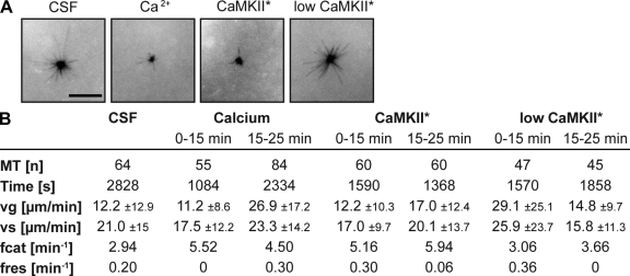
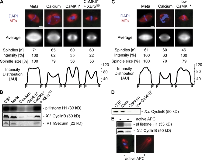
Altered spindle microtubule dynamics at anaphase onset are the basis for chromosome segregation. In Xenopus laevis egg extracts, increasing free calcium levels and subsequently rising calcium-calmodulin-dependent kinase II (CaMKII) activity promote a release from meiosis II arrest and reentry into anaphase. CaMKII induces the activation of the anaphase-promoting complex/cyclosome (APC/C), which destines securin and cyclin B for degradation to allow chromosome separation and mitotic exit. In this study, we investigated the calcium-dependent signal responsible for microtubule depolymerization at anaphase onset after release from meiotic arrest in Xenopus egg extracts. Using Ran-guanosine triphosphate-mediated microtubule assemblies and quantitative analysis of complete spindles, we demonstrate that CaMKII triggers anaphase microtubule depolymerization. A CaMKII-induced twofold increase in microtubule catastrophe rates can explain reduced microtubule stability. However, calcium or constitutively active CaMKII promotes microtubule destabilization even upon APC/C inhibition and in the presence of high cyclin-dependent kinase 1 activity. Therefore, our data demonstrate that CaMKII turns on parallel pathways to activate the APC/C and to induce microtubule depolymerization at meiotic anaphase onset.


Figures

Figure 1.
Figure 1.
Changes in microtubule stability from metaphase to anaphase in Xenopus egg extracts. (A) Calcium was added to CSF-arrested Xenopus egg extracts (0 min) in the presence of cyclinBΔ90, and samples were withdrawn at the indicated times to determine the activity of Cdk1 toward histone H1 (pHistone H1, top) and the amounts of endogenous cyclin B (X.I. cyclin B, bottom). (B) Microtubules or spindles were assembled in metaphase-arrested extracts (left), calcium was added in the presence of cyclinBΔ90 (right), and microtubules were visualized by direct fluorescence of added Cy3-tubulin. Sperm nuclei, RanQ69L, or chromatin beads were used as a nucleating source. (C) Immunoprecipitation using anti-TPX2 antibodies from metaphase or anaphase extracts. Immunocomplexes were eluted with high salt, and aurora A/Eg2 was determined by immunoblotting. (D) The microtubule assembly assay is as described in B; purified human centrosomes were used as a nucleating source. Cycled, microtubule asters were preassembled in metaphase before calcium addition; Meta, metaphase; Ana, anaphase. Bars, 10 μm.
Figure 2.
Figure 2.
Correlation of Cdk1 activity and changes in microtubule stability at metaphase to anaphase transition. (A) CSF-arrested egg extracts were incubated with calcium and cyclinBΔ90 (Ana) or CGP74514A (CGP). Ran-GTP–mediated microtubule assemblies were visualized by direct fluorescence after the addition of Cy3-labeled tubulin (top), and Cdk1 activities were determined by a histone H1 kinase activity assay (bottom). (B–D) CSF-arrested egg extracts (Meta or M) were triggered to go into anaphase by calcium addition in the presence of increasing cyclinBΔ90 concentrations. Ran-GTP–induced metaphase microtubule assemblies were visualized (B) and quantified relative to metaphase as indicated (C, black bars). In parallel, the Cdk1 kinase activity was measured using histone H1 phosphorylation (C, gray bars). (D) Anaphase was first induced with calcium in the presence of 80 nM cyclinBΔ90, increasing amounts of cyclinBΔ90 were added, and the capacity of Ran-GTP to still induce microtubule assembly was tested. These assemblies were quantified relative to metaphase (black bars), and the Cdk1 kinase activity was measured using histone H1 phosphorylation (gray bars). Error bars represent SD from three independent experiments. Ana, anaphase; Meta, metaphase. Bars, 5 μm.
Figure 3.
Figure 3.
APC/C activation is not required for changes in microtubule stability. (A) CSF extracts containing Cy3-tubulin and in vitro translated exogenously added securin were preincubated for 10 min in the presence of Ran-GTP to preassemble microtubules. Reactions were treated with buffer (Meta), calcium and cyclinBΔ90 (Ana), calcium (Inter), XErpND, or calcium and XErpND (XErpND + Ca2+). Microtubule structures were visualized by direct fluorescence of Cy3-tubulin. (B) Quantification of microtubule assemblies counted at the 40-min time point imaged in A. Error bars represent SD from three independent experiments; metaphase was set to 100%. (C) Amounts of cyclin B (endogenous, X.l. cyclin B; exogenously added [Δ90], H.s. cyclin B) were determined by immunoblotting, and amounts of exogenously added in vitro translated (IVT) securin were determined by autoradiography; Cdk1 activities were measured by histone H1 phosphorylation (pHistone H1). Meta, metaphase; Ana, anaphase; Inter, interphase. Bar, 5 μm.
Figure 4.
Figure 4.
CaMKII induces microtubule instability in anaphase. CSF extracts containing Cy3-tubulin and in vitro translated securin were preincubated for 10 min in the presence of RanQ69L to preassemble microtubules. (A) Reactions were treated with constitutively active CaMKII (CaMKII*), the catalytically inactive mutant (CaMKII*K42A), or CaMKII* together with cyclinBΔ90 (CaMKII* + Δ90) or XErpND (CamKII* + XErpND) as indicated. Microtubule assemblies were visualized by direct fluorescence. (B) Quantification of microtubule assemblies counted in A after 40 min. Error bars represent SD from three independent experiments; metaphase was set to 100%. (C) Reactions were performed as in A; the amounts of endogenous cyclin B (X.l. cyclin B) were determined by immunoblotting, and the amounts of exogenously added in vitro translated (IVT) securin were determined by autoradiography; Cdk1 activities were measured by histone H1 phosphorylation (pHistone H1). (D) Inhibition of endogenous CaMKII activity; CSF extracts containing Cy3-tubulin and RanQ69L were left untreated (Meta) or incubated with calcium in the absence (−) or presence (+) of the CaMKII inhibitor IINtide. (left) Microtubule structures were visualized by direct fluorescence of Cy3-tubulin. (right) Cyclin B (X.l. cyclin B) and Cdk1 activity (pHistone H1) were determined. (D, bottom) Reactions were chased using CaMKII* for the indicated time points and were visualized. Bars, 5 μm.
Figure 5.
Figure 5.
Dynamic measurements of centrosomally nucleated microtubules in metaphase or anaphase. CSF-arrested egg extracts were supplemented with Cy3-tubulin and purified human centrosomes and were triggered to enter anaphase as indicated. (A) Microtubules were visualized by time-lapse microscopy in 2-s intervals, and microtubules were tracked manually. Still images (after 15 min) are shown. (B) Parameters of dynamic instability were determined from data gained by time-lapse imaging using a Matlab macro. MT, microtubules; vg, growth rate; vs, shrinkage rate; fcat, catastrophe frequency; fres, rescue frequency. Bar, 10 μm.
Figure 6.
Figure 6.
CaMKII triggers spindle disassembly in anaphase independently of APC/C activation. Spindles were preassembled in CSF extracts (Meta), and anaphase was induced by the addition of calcium or CaMKII* in the absence or presence of XErpND. (A) Spindles were visualized by direct fluorescence of Cy3-tubulin (red) and DAPI (blue) after 20 min. Out of n imaged spindles, the mean overall fluorescence intensity was determined, and an average image was generated using the Matlab macro. The pole (P) to pole distance (spindle size) was measured as well as the corresponding mean fluorescence distribution plotted along the pole to pole axis in a 1.5-μm-wide area (intensity distribution; see blue lines in average images). Intensity and spindle size are indicated in relative terms (percentage). (B) Immunoblots were used to determine the amounts of cyclin B, an autoradiograph was used to monitor amounts of exogenously added in vitro translated (IVT) securin, and an autoradiograph displaying a histone H1 kinase assay was used to determine Cdk1 activity under the conditions used in A. The black line indicates that intervening lanes have been spliced out. (C) Anaphase was induced in preassembled spindles by low doses of CaMKII* in the presence of cyclinBΔ90, and samples were fixed after 40 min. Spindles were analyzed as in A. (D) Determination of cyclin B by immunoblotting under the conditions used in C. (E) Spindles were first treated with CaMKII* and XErpND and mixed with extracts treated without (−) or with (+) CaMKII* to activate APC/C. (top) Immunoblotting was used to determine the amounts of cyclin B and histone H1 kinase assay to measure the Cdk1 activity. (bottom) Representative fluorescence images of structures after 40 min. Tubulin, red; DAPI (DNA), blue. MT, microtubules; Meta, extracts after spindle assembly. Bars, 5 μm.

Similar articles

Cited by

References

    1. Blower, M.D., M. Nachury, R. Heald, and K. Weis. 2005. A Rae1-containing ribonucleoprotein complex is required for mitotic spindle assembly. Cell. 121:223–234. - PubMed
    1. Busa, W.B., and R. Nuccitelli. 1985. An elevated free cytosolic Ca2+ wave follows fertilization in eggs of the frog, Xenopus laevis. J. Cell Biol. 100:1325–1329. - PMC - PubMed
    1. Carazo-Salas, R.E., G. Guarguaglini, O.J. Gruss, A. Segref, E. Karsenti, and I.W. Mattaj. 1999. Generation of GTP-bound Ran by RCC1 is required for chromatin-induced mitotic spindle formation. Nature. 400:178–181. - PubMed
    1. Carazo-Salas, R.E., O.J. Gruss, I.W. Mattaj, and E. Karsenti. 2001. Ran-GTP coordinates regulation of microtubule nucleation and dynamics during mitotic-spindle assembly. Nat. Cell Biol. 3:228–234. - PubMed
    1. Cassimeris, L., and R.V. Skibbens. 2003. Regulated assembly of the mitotic spindle: a perspective from two ends. Curr. Issues Mol. Biol. 5:99–112. - PubMed

Publication types

Substances