. 2008 Dec 22;205(13):3007-18.
doi: 10.1084/jem.20081165. Epub 2008 Dec 8.

Induction of inflammatory and immune responses by HMGB1-nucleosome complexes: implications for the pathogenesis of SLE

Affiliations

Induction of inflammatory and immune responses by HMGB1-nucleosome complexes: implications for the pathogenesis of SLE

Vilma Urbonaviciute et al. J Exp Med. .

Abstract

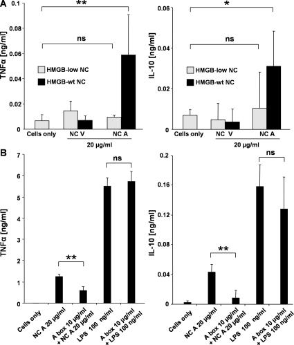
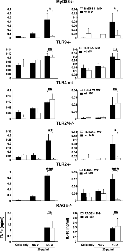
Autoantibodies against double-stranded DNA (dsDNA) and nucleosomes represent a hallmark of systemic lupus erythematosus (SLE). However, the mechanisms involved in breaking the immunological tolerance against these poorly immunogenic nuclear components are not fully understood. Impaired phagocytosis of apoptotic cells with consecutive release of nuclear antigens may contribute to the immune pathogenesis. The architectural chromosomal protein and proinflammatory mediator high mobility group box protein 1 (HMGB1) is tightly attached to the chromatin of apoptotic cells. We demonstrate that HMGB1 remains bound to nucleosomes released from late apoptotic cells in vitro. HMGB1-nucleosome complexes were also detected in plasma from SLE patients. HMGB1-containing nucleosomes from apoptotic cells induced secretion of interleukin (IL) 1beta, IL-6, IL-10, and tumor necrosis factor (TNF) alpha and expression of costimulatory molecules in macrophages and dendritic cells (DC), respectively. Neither HMGB1-free nucleosomes from viable cells nor nucleosomes from apoptotic cells lacking HMGB1 induced cytokine production or DC activation. HMGB1-containing nucleosomes from apoptotic cells induced anti-dsDNA and antihistone IgG responses in a Toll-like receptor (TLR) 2-dependent manner, whereas nucleosomes from living cells did not. In conclusion, HMGB1-nucleosome complexes activate antigen presenting cells and, thereby, may crucially contribute to the pathogenesis of SLE via breaking the immunological tolerance against nucleosomes/dsDNA.


Figures

Figure 1.
Figure 1.
HMGB1 is associated with nucleosomes spontaneously released during secondary necrosis. For induction of secondary necrosis (SN), Jurkat cells were treated with 2 μM staurosporine for 48 h. Primary necrosis was induced by heating cells at 56°C for 30 min followed by incubation for an additional hour at 37°C. Supernatants from apoptotic (top) and necrotic (bottom) Jurkat cells were collected and subjected to immune precipitations with anti-HMGB1, anti-dsDNA, antihistone H3, antihistone H2B, and antihistones H2A/H4 as well as with appropriate isotype control antibodies. Immune-precipitated material was then fractionated by SDS-PAGE on 12% polyacrylamide gel. Western blot analysis was performed using polyclonal anti-HMGB1 antibodies. WB, Western blotting; IP, immunoprecipitation. These experiments have been performed four times with virtually identical results.
Figure 2.
Figure 2.
Nucleosomes circulating in the blood of patients with SLE are complexed with HMGB1. (A) Circulating immune complexes in the blood of patients with SLE contain HMGB1. Immune complexes from serum samples of patients with SLE and rheumatoid arthritis (RA) as well as from healthy persons (NHD, normal healthy donors) were precipitated by 1.5 or 2% of polyethylene glycol (PEG). The precipitates were dissolved in SDS sample buffer and analyzed by Western blotting using polyclonal anti-HMGB1 antibodies. (B) Antihistone and anti-dsDNA antibodies coimmunoprecipitate HMGB1 from sera of patients with SLE. Serum samples obtained from patients with SLE and normal healthy donors were subjected to coimmunoprecipitation using antihistone and anti-dsDNA antibodies covalently linked to protein G Sepharose. Sepharose beads alone or linked to isotype control antibodies served as negative controls. The precipitates were separated by reducing SDS-PAGE on a 12.5% polyacrylamide gel followed by Western blot analysis using polyclonal anti-HMGB1 antibodies.
Figure 3.
Figure 3.
HMGB1 cofractionates with nucleosomes from apoptotic cells. For induction of apoptosis, Jurkat cells were treated with 1 μM staurosporine for 8 h. Necrosis was induced by heat treatment at 56°C for 30 min. Nuclei isolated from viable (A), necrotic (B), and apoptotic (C) Jurkat cells were digested with micrococcal nuclease, lysed, and fractionated by sucrose gradient ultracentrifugation. Eight fractions were collected in each gradient from top (1) to bottom (8), and individual fractions were analyzed by 1.5% agarose gel electrophoresis in the presence of 1% SDS. HMGB1 was detected by Western blotting. (D) Characterization of mononucleosomes. 10 μg of total protein of mononucleosomal fraction 4 purified from necrotic (lane 1), viable (lane 2), and apoptotic (lane 3) Jurkat cells was subjected to a 15% SDS-PAGE. The proteins were visualized by Coomassie blue staining.
Figure 4.
Figure 4.
HMGB1-containing nucleosomes from apoptotic cells induce secretion of cytokines by human monocyte-derived macrophages. Human macrophages were cultured in the absence or presence of 20 μg/ml of nucleosomes purified from viable (NC V), apoptotic (NC A), and necrotic cells (NC N) or diluent (PBS). Untreated human macrophages served as controls. Cell culture supernatants were harvested after 24 h and cytokine concentrations were determined. One representative of three independent experiments is shown. Mean values and SD were calculated from triplicates. Student's t test was used for statistical analysis. **, P ≤ 0.01; *, P ≤ 0.05.
Figure 5.
Figure 5.
HMGB1 contributes to the proinflammatory activity of apoptotic cell–derived nucleosomes. (A) Nucleosomes isolated from apoptotic WT, but not from HMGBlow MEF, stimulated human monocyte-derived macrophages to release TNF-α and IL-10. Macrophages were incubated with 20 μg/ml of nucleosomes purified from viable (NC V) and apoptotic (NC A) WT or HMGBlow MEF. Untreated macrophages served as control. After 24 h, concentrations of TNF-α and IL-10 were measured by ELISA. (B) Apoptotic nucleosome-induced cytokine release was suppressed by the antagonistic A box domain of HMGB1. Macrophages were stimulated either with 20 μg/ml of nucleosomes purified from apoptotic cells or 100 ng/ml LPS in the absence or presence of 10 μg/ml of the recombinant A box fragment. After incubation for 24 h, cell culture supernatants were harvested and concentrations of the cytokines TNF-α and IL-10 were measured by ELISA. Untreated macrophages served as a control. One representative example of three experiments is shown. Mean values and SD were calculated from triplicates. Student's t test was used for statistical analysis. **, P ≤ 0.01; *, P ≤ 0.05.
Figure 6.
Figure 6.
Apoptotic nucleosome–induced cytokine release by macrophages is dependent on MyD88 and TLR2 but not on TLR4, TLR9, and RAGE. Thioglycollate-elicited peritoneal macrophages were obtained from WT, MyD88-, TLR9-, TLR2/4-, TLR2-, and RAGE-deficient or TLR4 mutant mice and incubated with 20 μg/ml of nucleosomes purified from viable (NC V) and apoptotic (NC A) Jurkat cells for 24 h. Untreated macrophages served as a control. Concentrations of TNF-α and IL-10 were measured by ELISA. Mean values and SD were calculated from triplicates. Student's t test was used for statistical analysis. ***, P ≤ 0.001; **, P ≤ 0.01; *, P ≤ 0.05.
Figure 7.
Figure 7.
Nucleosomes induce maturation of DC. (A) Monocyte-derived DC were cultured in the absence or presence of 20 μg/ml of nucleosomes purified from viable (NC V) or apoptotic (NC A) Jurkat cells. After incubation for 48 h, expression of MHC class II, CD86, and CD83 molecules was assessed by flow cytometry. The results are expressed as mean fluorescence intensity (MFI). On the left, bar graphs show means and SD of triplicates. On the right, corresponding representative histograms are shown. Gray line, unstimulated DC; black line, DC exposed to nucleosomes from viable cells; filled gray, DC exposed to nucleosomes from apoptotic cells. (B) DC exposed to nucleosomes from apoptotic cells are more potent stimulators of allogeneic T cells than DC exposed to nucleosomes from viable cells. Immature DC were incubated for 48 h with nucleosomes purified from viable or apoptotic cells or LPS as positive control. DC were then cocultured with allogeneic T cells at a DC/T cell ratio of 1:100. T cell proliferation was assessed by measuring the amount of 3H thymidine incorporation. Mean values and SD were calculated from triplicates. Student's t test was used for statistical analysis. **, P ≤ 0.01. (C) HMGB1 contributes to the apoptotic nucleosome–induced DC activation. Human monocyte-derived DC were left untreated or incubated with 20 μg/ml of nucleosomes purified from viable or apoptotic WT or HMGBlow MEF. After 48 h, expression of CD83 and CD86 on the surface of DC was determined by flow cytometry. The results are expressed as mean fluorescence intensity. Student's t test was used for statistical analysis. **, P ≤ 0.01; *, P ≤ 0.05. Experiments were repeated three times using DC generated from different donors.
Figure 8.
Figure 8.
Increased immunogenicity of nucleosomes derived from apoptotic cells and involvement of TLR2 in the autoimmune response. (A) Groups of five BALB/c mice each were i.v. immunized three times with 50 μg of purified nucleosomes from viable and apoptotic Jurkat cells in intervals of 3 wk. A control group received PBS. Sera were collected before the first immunization and 3 wk after each immunization. Concentrations of IgG antibodies against nucleosomes, ssDNA, dsDNA, and histones were determined by ELISA. All sera were tested in the same assay. Mean values of the OD and SD are shown. Statistical analysis was performed using the nonparametric Mann-Whitney U test for unpaired samples. **, P ≤ 0.01; *, P ≤ 0.05. (B) To evaluate the requirement of TLR2 for the production of autoantibodies in vivo, groups of TLR2−/−, TLR2/4−/−, and C57BL/6 mice were i.v. injected with 75 μg of purified nucleosomes from apoptotic cells in intervals of 2 wk. 2 wk after the third immunization, anti-dsDNA and antihistone IgG antibodies were quantified in serum samples by ELISA. All sera were tested in the same assay. Bars indicate the mean values. Statistical analyses were performed using the Student's t test for unpaired samples. *, P ≤ 0.05.

References

    1. Tan, E.M., P.H. Schur, R.I. Carr, and H.G. Kunkel. 1966. Deoxybonucleic acid (DNA) and antibodies to DNA in the serum of patients with systemic lupus erythematosus. J. Clin. Invest. 45:1732–1740. - PMC - PubMed
    1. Hahn, B.H. 1998. Antibodies to DNA. N. Engl. J. Med. 338:1359–1368. - PubMed
    1. Winfield, J.B., I. Faiferman, and D. Koffler. 1977. Avidity of anti-DNA antibodies in serum and IgG glomerular eluates from patients with systemic lupus erythematosus. Association of high avidity antinative DNA antibody with glomerulonephritis. J. Clin. Invest. 59:90–96. - PMC - PubMed
    1. Ehrenstein, M.R., D.R. Katz, M.H. Griffiths, L. Papadaki, T.H. Winkler, J.R. Kalden, and D.A. Isenberg. 1995. Human IgG anti-DNA antibodies deposit in kidneys and induce proteinuria in SCID mice. Kidney Int. 48:705–711. - PubMed
    1. Thomas, J.O., and A.A. Travers. 2001. HMG1 and 2, and related ‘architectural’ DNA-binding proteins. Trends Biochem. Sci. 26:167–174. - PubMed

Publication types

MeSH terms