

Review
doi: 10.1001/archneur.65.12.1571.
Transcranial direct current stimulation in stroke recovery
Affiliations
- PMID: 19064743
- PMCID: PMC2779259
- DOI: 10.1001/archneur.65.12.1571
Item in Clipboard
Review
Transcranial direct current stimulation in stroke recovery
Arch Neurol.
2008 Dec.
Abstract
Transcranial direct current stimulation (TDCS) is an emerging technique of noninvasive brain stimulation that has been found useful in examining cortical function in healthy subjects and in facilitating treatments of various neurologic disorders. A better understanding of adaptive and maladaptive poststroke neuroplasticity and its modulation through noninvasive brain stimulation has opened up experimental treatment options using TDCS for patients recovering from stroke. We review the role of TDCS as a facilitator of stroke recovery, the different modes of TDCS, and the potential mechanisms underlying the neural effects of TDCS.
Figures
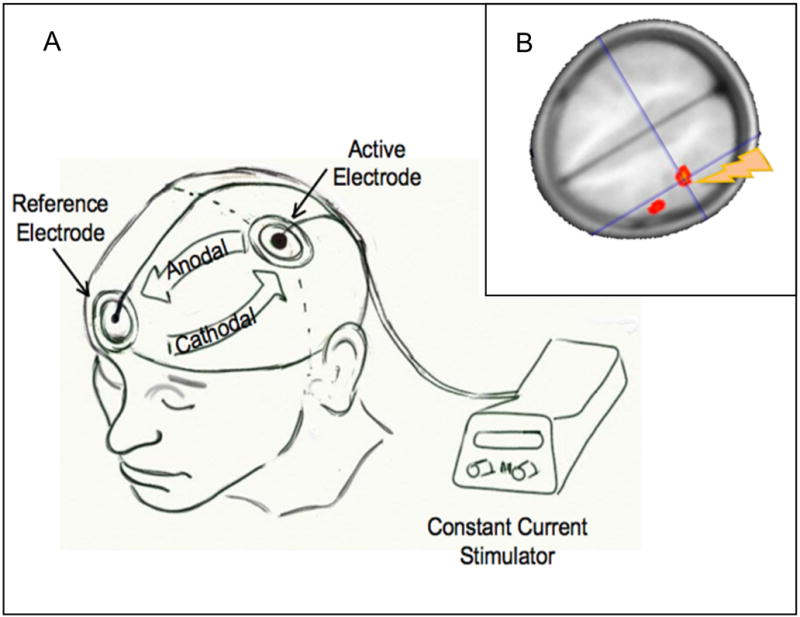
A) This drawing shows the TDCS setup using a mobile, battery-operated direct current stimulator connected with two electrodes. One electrode (active) is positioned over C3 (corresponding to the precentral gyrus) and the reference electrode is positioned over the contralateral supraorbital region. If current flows from C3 to the supraorbital region, then the tissue underlying C3 is subjected to anodal (increase in excitability) stimulation. If current is reversed, then the tissue underlying C3 is subjected to cathodal (decrease in excitability) stimulation; B) Regional cerebral blood increases in the motor region underlying the electrode positioned over C3 after anodal stimulation. Regional cerebral blood was determined using a non-invasive arterial spin-labeling technique.
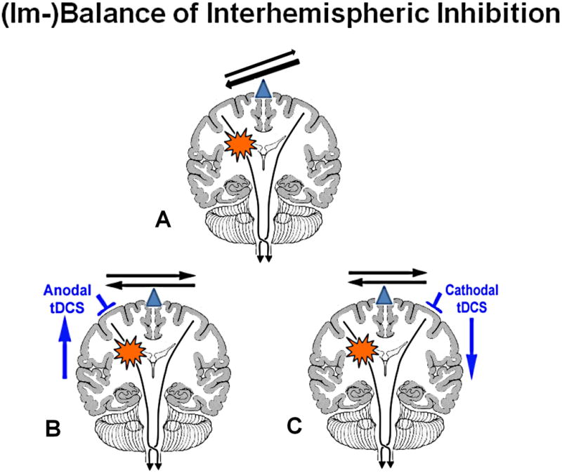
The balance of interhemispheric inhibition becomes disrupted after a stroke (A). This leaves the healthy hemisphere in a position that it could exert too much of an unopposed/imbalanced inhibitory influence onto the lesional hemisphere and possibly interfere in the recovery process of the affected hemisphere. There are two possible ways to ameliorate this imbalance: either one upregulates the excitability in the affected (lesional) hemisphere (B) or one downregulates the excitability in the unaffected (normal) hemisphere (C).
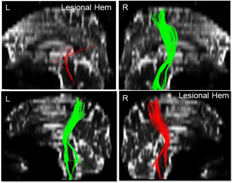
This picture shows two patients with their representative corticospinal tract (CST) fibers that originate from the white matter underlying the precentral gyrus and travel through the internal capsule into the brainstem. The CST of the lesional hemispheres differs between both patients. Patient #1 shows a severely reduced number of fibers that don’t seem to originate from the dorsal part of the motor region (hand/arm region of the precentral gyrus), but still show a path through the posterior limb of the internal capsule into the brainstem while patient #2 has a very mild reduction in the number of CST fibers on the lesional hemisphere, but otherwise shows a similar origin and descent of the CST between the lesional and normal hemisphere. The improvement after TDCS stimulation was pronounced in the patient with intact pyramidal tract, but only minimal in the patient with the disrupted pyramidal tract.
References
-
- Priori A. Brain polarization in humans: a reappraisal of an old tool for prolonged non-invasive modulation of brain excitability. Clin Neurophysiol. 2003;114(4):589–95. - PubMed
-
- Gross CG. The discovery of motor cortex and its background. J Hist Neurosci. 2007;16(3):320–31. - PubMed
-
- Gilula MF, Kirsch DL. Cranial Electrotherapy Stimulation review: A Safer Alternative to Pyschopharmaceuticals in the Treatment of Depression. J Neurotherapy. 2005;9(2):7–26.
-
- Islam N, Aftabuddin M, Moriwaki A, Hattori Y, et al. Increase in the calcium level following anodal polarization in the rat brain. Brain Res. 1995;684(2):206–8. - PubMed
Publication types
MeSH terms
Grants and funding
LinkOut - more resources
Full Text Sources
Other Literature Sources
Medical
