

Hippocampal volumes, proton magnetic resonance spectroscopy metabolites, and cerebrovascular disease in mild cognitive impairment subtypes
- PMID: 19064749
- PMCID: PMC2743393
- DOI: 10.1001/archneur.65.12.1621
Hippocampal volumes, proton magnetic resonance spectroscopy metabolites, and cerebrovascular disease in mild cognitive impairment subtypes
Abstract
Background: Although a majority of patients with amnestic mild cognitive impairment (aMCI) progress to Alzheimer disease, the natural history of nonamnestic MCI (naMCI) is less clear. Noninvasive imaging surrogates for underlying pathological findings in MCI would be clinically useful for identifying patients who may benefit from disease-specific treatments at the prodromal stage of dementia.
Objective: To determine the characteristic magnetic resonance imaging (MRI) and proton MR spectroscopy (1H MRS) profiles of MCI subtypes.
Design: Case-control study.
Setting: Community-based sample at a tertiary referral center.
Patients: Ninety-one patients with single-domain aMCI, 32 patients with multiple-domain aMCI, 20 patients with single- or multiple-domain naMCI, and 100 cognitively normal elderly subjects frequency-matched by age and sex.
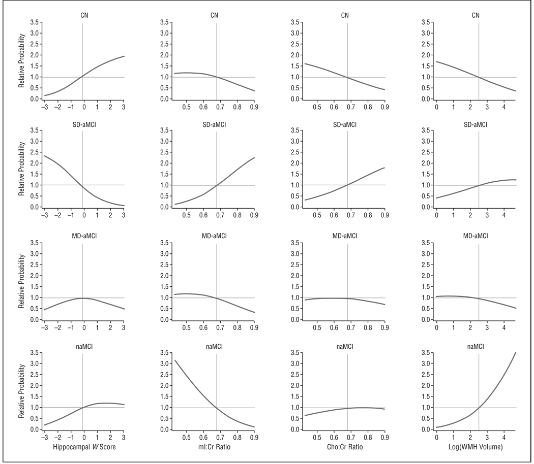
Main outcome measures: Posterior cingulate gyrus 1H MRS metabolite ratios, hippocampal volumes, and cerebrovascular disease on MRI.
Results: Patients with single-domain aMCI were characterized by small hippocampal volumes and elevated ratios of myo-inositol to creatine levels. Patients with naMCI on average had normal hippocampal volumes and 1H MRS metabolite ratios, but a greater proportion (3 of 20 patients [15%]) had cortical infarctions compared with patients with single-domain aMCI (6 of 91 [7%]). For characterization of MCI subtypes, 1H MRS and structural MRI findings were complementary.
Conclusions: The MRI and 1H MRS findings in single-domain aMCI are consistent with a pattern similar to that of Alzheimer disease. Absence of this pattern on average in patients with naMCI suggests that cerebrovascular disease and other neurodegenerative diseases may be contributing to the cognitive impairment in many individuals with naMCI.
Figures
References
-
- Petersen RC. Mild cognitive impairment. Continuum. 2004;10(1):9–28.
-
- Fischer P, Jungwirth S, Zehetmayer S, et al. Conversion from subtypes of mild cognitive impairment to Alzheimer dementia. Neurology. 2007;68(4):288–291. - PubMed
-
- Flicker C, Ferris SH, Reisberg B. Mild cognitive impairment in the elderly: predictors of dementia. Neurology. 1991;41(7):1006–1009. - PubMed
-
- Morris JC, Storandt M, Miller JP, et al. Mild cognitive impairment represents early-stage Alzheimer disease. Arch Neurol. 2001;58(3):397–405. - PubMed
-
- Busse A, Hensel A, Guhne U, Angermeyer MC, Riedel-Heller SG. Mild cognitive impairment: long-term course of four clinical subtypes. Neurology. 2006;67(12):2176–2185. - PubMed
