. 2009 Jul;14(7):719-27.
doi: 10.1038/mp.2008.130. Epub 2008 Dec 9.

Targeted disruption of serine racemase affects glutamatergic neurotransmission and behavior

Affiliations

Targeted disruption of serine racemase affects glutamatergic neurotransmission and behavior

A C Basu et al. Mol Psychiatry. 2009 Jul.

Erratum in

  • Mol Psychiatry. 2010 Nov;15(11):1122

Abstract

A subset of glutamate receptors that are specifically sensitive to the glutamate analog N-methyl-D-aspartate (NMDA) are molecular coincidence detectors, necessary for activity-dependent processes of neurodevelopment and in sensory and cognitive functions. The activity of these receptors is modulated by the endogenous amino acid D-serine, but the extent to which D-serine is necessary for the normal development and function of the mammalian nervous system was previously unknown. Decreased signaling at NMDA receptors has been implicated in the pathophysiology of schizophrenia based on pharmacological evidence, and several human genes related to D-serine metabolism and glutamatergic neurotransmission have been implicated in the etiology of schizophrenia. Here we show that genetically modified mice lacking the ability to produce D-serine endogenously have profoundly altered glutamatergic neurotransmission, and relatively subtle but significant behavioral abnormalities that reflect hyperactivity and impaired spatial memory, and that are consistent with elevated anxiety.


Figures

Figure 1
Figure 1
The serine racemase exon 1 knockout (SR−/−) mouse model. (a) Exon 1 of the SR gene was targeted for deletion by insertion of a loxP sequence and a loxP-flanked PGK-Neo neomycin resistance sequence at upstream and downstream restriction sites respectively. Breeding of animals carrying the targeted locus with animals that constitutively expressed Cre protein produced offspring carrying desired constructs. (b) Western blot of whole brain protein extracts revealed a decrease in SR protein in SR+/− animals and an absence of SR protein in SR−/− animals. (c) SR protein expression was evident throughout the cortex, striatum, and hippocampus of WT mice. (d) SR protein expression was undetectable in SR−/− mice by immunohistochemical analysis. Scale bar represents 2 mm.
Figure 2
Figure 2
Changes in NMDAR-mediated physiology in SR mutant mice. Values are shown and stated as Mean ± SEM. P-values less than 0.05, indicated by asterisks, are of comparisons to wild type using one-tailed Student’s t test. (a) NMDAR EPSCs take longer to decay in SR mutants. (b) Time constant of decay (tau) of NMDAR EPSCs is higher in SR−/− mice (WT: 124.32 ± 8.72 ms, n=18; SR+/− : 144.31 ± 11.01 ms, n=16, p < 0.05 ; SR−/− : 157.38 ± 8.67 ms, n=16, p < 0.05). (c) Baseline amplitudes (black) of NMDAR EPSCs of WT, SR+/− and SR−/− were enhanced by application of 10 μM D-serine (blue) or 100 nM NFPS (green). (d) Amplitude increase of NMDAR EPSCs induced by 10 μM D-serine was higher in SR +/− and SR −/− compared to WT mice (WT: 41.29 ± 10.93% , n=4; SR+/− : 81.03 ±11.21% , n=5, p < 0.05; SR−/− : 93.19 ± 2.24%, n=5, p < 0.05). ) Amplitude increase of NMDAR EPSCs induced by 100 nM NFPS was higher in SR −/− compared to WT mice (WT: 40.68 ± 4.04 %, n=4; SR+/−: 62.11 ± 15.57%, n=5; SR−/− : 69.19 ±11.73%, n=6, p < 0.05. (e) Biotin-switch assay showed that SR−/− mice have ~70% reduced S-nitrosylation of proteins (n=4 p<0.001) (f) Using a pairing protocol to induce LTP, no significant changes were observed in the amplitude of the EPSC in SR−/− (○, 23.72 ± 11.75%, n = 10) while the same protocol induced an increase in the amplitude of EPSC in WT mice (△, 104.96 ± 22.5%, n = 5). 10 μM D-serine in the bath solution 10 min prior to the application of the protocol restored LTP in slices from SR−/− mice (●, 93.71 + 6.83%, n = 5).
Figure 3
Figure 3
Motor activity and startle in SR−/− mice. (a) SR−/− mice showed no significant deficit in motor coordination or learning in a three-day rotarod task. (b) SR−/− male mice traversed more distance and (c) exhibited more vertical behavior (rearing and jumping) than WT controls in a three-day locomotor activity assay. (d) SR−/− female mice spent less time in the center zone of the activity chamber on the first day of the activity assay. (e) SR−/− showed no deficit in prepulse inhibition (PPI) of the acoustic startle response (ASR). (f) All experimental groups showed habituation of the ASR, but SR−/− female mice showed elevated startle reactivity. Values are shown as Mean ± SEM. Male WT: blue open squares; male SR−/−: blue closed squares; female WT: red open circles; female SR−/−: red closed circles; n= 15 per group.
Figure 4
Figure 4
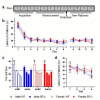
Spatial learning and memory in SR−/− mice. (a) Mice were trained to learn the location of a hidden platform for eight days, subjected to a probe test on the eighth day, and then tested with new platform positions on each day for four days. (b) SR−/− mice matched WT performance during as assessed by latency to find the hidden platform. (c) The probe test revealed a deficit in spatial reference memory in SR−/− males. For each experimental group, the bars shown represent the percent time spent in the target quadrant (formerly containing the platform), an adjacent quadrant, the opposite quadrant, and the other adjacent quadrant of the maze. (d) There was no significant difference between SR−/− and WT mice in the trial-to-trial learning of new platform positions as assessed by latency to platform. Values shown are mean latencies for trials 1-4 from days 9-12. Values are shown as Mean ± SEM. Male WT: blue open squares; male SR−/−: blue closed squares; female WT: red open circles; female SR−/−: red closed circles; n= 15 per group.

References

    1. Johnson JW, Ascher P. Glycine potentiates the NMDA response in cultured mouse brain neurons. Nature. 1987;325:529–531. - PubMed
    1. Kleckner NW, Dingledine R. Requirement for glycine in activation of NMDA receptors expressed in Xenopus Oocytes. Science. 1988;241:835–837. - PubMed
    1. Fadda E, Danysz W, Wroblewski JT, Costa E. Glycine and D-serine increase the affinity of N-methyl-D-aspartate sensitive glutamate binding sites in rat brain synaptic membranes. Neuropharmacology. 1988;27:1183–1185. - PubMed
    1. Matsui T, Sekiguchi M, Hashimoto A, Tomita U, Nishikawa T, Wada K. Functional comparison of D-serine and glycine in rodents: the effect on cloned NMDA receptors and the extracellular concentration. J Neurochem. 1995;65:454–458. - PubMed
    1. Mothet JP, Parent AT, Wolosker H, Brady RO, Jr, Linden DJ, Ferris CD, et al. D-serine is an endogenous ligand for the glycine site of the N-methyl-D-aspartate receptor. Proc Natl Acad Sci USA. 2000;97:4926–4931. - PMC - PubMed

Publication types

MeSH terms