. 2009 Jan;39(1):234-40.
doi: 10.1002/eji.200838772.

CD4+ T-cell development in a mouse expressing a transgenic TCR derived from a Treg

Affiliations

CD4+ T-cell development in a mouse expressing a transgenic TCR derived from a Treg

Richard J DiPaolo et al. Eur J Immunol. 2009 Jan.

Abstract

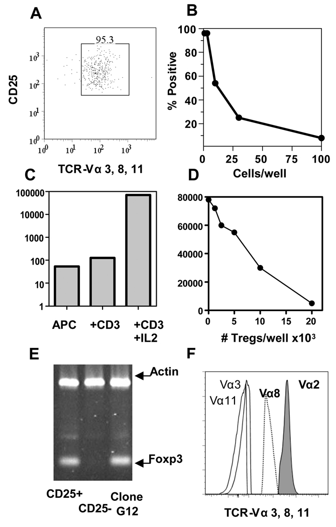
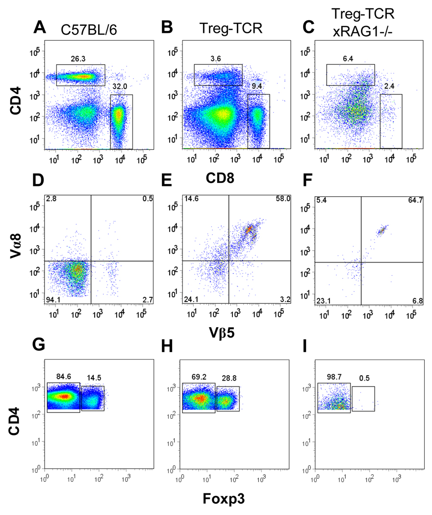
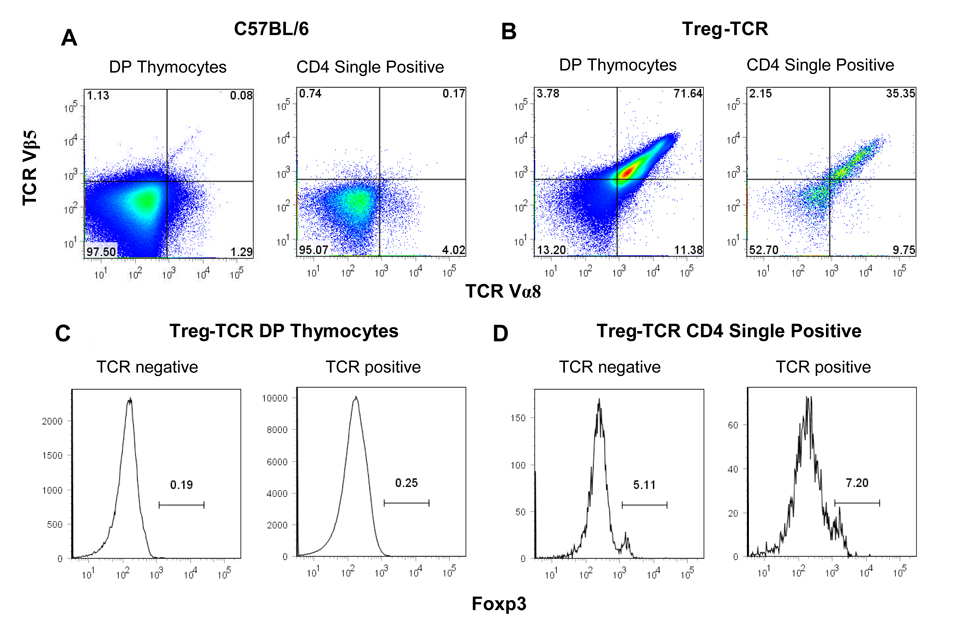
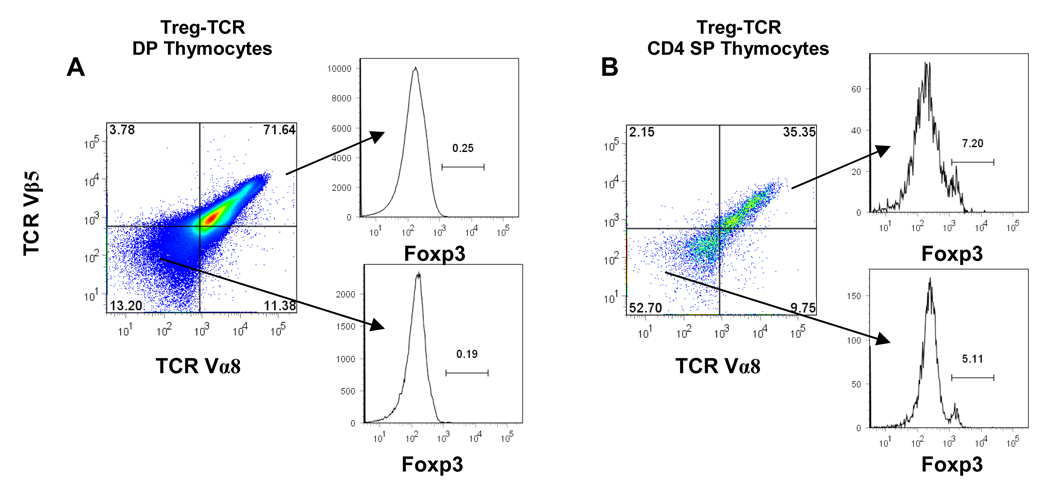
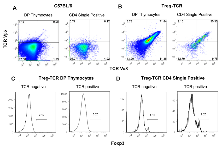
CD4(+)Foxp3(+) Treg maintain peripheral tolerance and influence immune responses to foreign antigens. The thymus is an important source of Treg, but controversy exists as to whether T cells are selected into the Treg lineage based on signals received through TCR specific for self-peptides. To examine the specificity of TCR expressed by Treg and its effect on CD4(+) T-cell development, we generated Treg-TCR transgenic mice. Deletion of >90% of CD4(+) T cells in RAG-sufficient mice, and nearly 100% deletion in RAG(-/-) mice expressing this TCR indicate that the TCR is specific for an unknown, naturally expressed peptide in the thymus. Deletion occurs late in development, suggesting this peptide is presented by APC in the thymic medulla. These studies are the first to describe the effects of expressing a Treg-TCR on CD4(+) T-cell development. The implications of our data for models of Treg selection are discussed.


Conflict of interest statement

The authors declare no financial or commercial conflict of interest.

Figures

Figure 1
Figure 1. Cloning Tregs and identifying Treg-TCRs
(A) >95% of the CD4+ T cells express CD25 and TCR Vα3, Vα8, and Vα11 after sorting lymph nodes cells from OTII TCR transgenic mice. (B) Results of plating the sorted cells in limiting dilution and scoring wells for T cell growth. (C) The proliferation of an individual clone, G12, to APCs alone, APCs + anti-CD3, and APCs + anti-CD3 + IL-2. (D) Clone (G12) was tested for the ability to suppress the proliferation of freshly isolated CD4+ T cells responding to anti-CD3. (E) Foxp3 expression by freshly isolated CD4+CD25+ T cells, CD4+CD25 T cells, and the Treg T cell clone G12. (F) The expression of TCR Vα8, and Vα11 was analyzed by flow cytometry on clone G12, along with Vα2 and Vα3 as negative controls.
Figure 2
Figure 2. Reduced CD4+Foxp3 and CD4+Foxp3+ T cells in the periphery of Treg-TCR transgenic mice
(A–C) The proportions of CD4+ and CD8+ T cell in the spleens of C57BL/6, Treg-TCR, and Treg-TCRxRAG1−/− transgenic mice. (D–F) Expression of TCR Vβ5 and TCR Vα8 on CD4+ gated T cells from the spleen. (G–H) Foxp3 expression on CD4+ gated T cells from the spleen. Total numbers of cells are shown in Table I. Results shown are representative of similar analysis performed on 7 to 10 individual mice in each group.
Figure 3
Figure 3. Deletion of CD4+Foxp3 and CD4+Foxp3+ thymocytes in Treg-TCR transgenic mice
The expression of CD4 and CD8 from (A) negative control littermates (C57BL/6), (B) Treg-TCR transgenic mice, and (C) Treg-TCRxRAG1−/− (D) Treg-TCRxOTII, and (E) OTII TCR transgenic mice. Expression of CD25 and Foxp3 on CD4+CD8 gated thymocytes from (E) negative control littermates, (F) Treg-TCR transgenic mice, (G) Treg-TCRxRAG1−/− mice, (H) Treg-TCRxOTII, and (I) OTII TCR transgenic mice. Total numbers of thymocytes are shown in Table I. Results are representative of similar analysis of between 4 and 10 individual mice in each group.
Figure 4
Figure 4. TCR and Foxp3 expression in CD4+CD8+ double positive (DP), and CD4 single positive (SP) thymocytes from Treg-TCR transgenic mice
The expression of the transgenic TCR Vα8 and TCR Vβ5 on DP gated thymocytes (A), and CD4 SP gated thymocytes (B) from Treg-TCR transgenic mice. Foxp3 expression is shown after gating on the TCR Vα8/Vβ5 high, intermediate, and low DP thymocytes (A) and CD4SP thymocytes (B). Results are represented of 7 individual mice in each group.

References

    1. Fontenot JD, Dooley JL, Farr AG, Rudensky AY. Developmental regulation of Foxp3 expression during ontogeny. J Exp Med. 2005;202:901–906. - PMC - PubMed
    1. Apostolou I, Sarukhan A, Klein L, von Boehmer H. Origin of regulatory T cells with known specificity for antigen. Nat Immunol. 2002;3:756–763. - PubMed
    1. Jordan MS, Boesteanu A, Reed AJ, Petrone AL, Holenbeck AE, Lerman MA, Naji A, Caton AJ. Thymic selection of CD4+CD25+ regulatory T cells induced by an agonist self-peptide. Nat Immunol. 2001;2:301–306. - PubMed
    1. Kawahata K, Misaki Y, Yamauchi M, Tsunekawa S, Setoguchi K, Miyazaki J, Yamamoto K. Generation of CD4(+)CD25(+) regulatory T cells from autoreactive T cells simultaneously with their negative selection in the thymus and from nonautoreactive T cells by endogenous TCR expression. J Immunol. 2002;168:4399–4405. - PubMed
    1. Larkin J, 3rd, Rankin AL, Picca CC, Riley MP, Jenks SA, Sant AJ, Caton AJ. CD4+CD25+ Regulatory T Cell Repertoire Formation Shaped by Differential Presentation of Peptides from a Self-Antigen. J Immunol. 2008;180:2149–2157. - PubMed

Publication types

Substances

LinkOut - more resources