Review
. 2008:640:234-51.
doi: 10.1007/978-0-387-09789-3_18.

Therapeutic blockade of T-cell antigen receptor signal transduction and costimulation in autoimmune disease

Affiliations
Review

Therapeutic blockade of T-cell antigen receptor signal transduction and costimulation in autoimmune disease

Joseph R Podojil et al. Adv Exp Med Biol. 2008.

Abstract

CD4+ T-cell-mediated autoimmune diseases are initiated and maintained by the presentation of self-antigen by antigen-presenting cells (APCs) to self-reactive CD4+ T-cells. According to the two-signal hypothesis, activation of a naive antigen-specific CD4+ T-cell requires stimulation of both the T-cell antigen receptor (signal 1) and costimulatory molecules such as CD28 (signal 2). To date, the majority of therapies for autoimmune diseases approved by the Food and Drug Administration primarily focus on the global inhibition of immune inflammatory activity. The goal of ongoing research in this field is to develop antigen-specific treatments which block the deleterious effects of self-reactive immune cell function while maintaining the ability of the immune system to clear nonself antigens. To this end, the signaling pathways involved in the induction of CD4+ T-cell anergy, as apposed to activation, are a topic of intense interest. This chapter discusses components of the CD4+ T-cell activation pathway that may serve as therapeutic targets for the treatment of autoimmune disease.


Figures

Figure 1
Figure 1. Clinical disease courses in multiple sclerosis (MS) and and its mouse model, experimental autoimmune encephalomyelitis (EAE)
The clinical disease course of MS is classified according to the characteristics and severity of disease progression over time. The most common disease course of MS is Relapsing-Remitting MS (RRMS). This disease course is characterized by a defined acute attack (increase in disability), which is followed by a full recovery and subsequent attacks over time. Secondary Progressive MS (SPMS) is similar to RRMS, but instead of full recovery during remission, residual deficit is maintained. SPMS is characterized by less recovery during remission following attacks and fewer attacks as the disease course switches from a relapsing-remitting disease course to a more progressive disease course. Primary Progressive MS (PPMS) is a disease course characterized by a progressive increase in disability over time in the absence of well-defined relapses and/or remissions. Progressive-Relapsing MS (PRMS) is the least common of the disease courses characterized by a progressive disability from the onset of disease. PRMS contains clear relapses in disease severity in the absence or presence of full recovery. In the SJL mouse, relapsing-remitting model of MS, i.e., R-EAE, the dominate spread epitope for each consecutive relapse is well characterized. During the acute phase of the disease the majority of activated CD4+ T cells are specific for the dominate proteolipid protein epitope, PLP139-151, used to induce disease. The dominate epitope during the primary relapse is PLP178-191 (intramolecular epitope spreading) and during the secondary relapse this epitope is MBP84-104 (intermolecular epitope spreading). In contrast, epitope spreading in the chronic disease model has been suggested, but the consecutive dominant epitopes have not been identified.
Figure 2
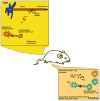
Figure 2. Epitope spreading
Animal models of multiple sclerosis (MS) have helped to identify putative mechanisms by which epitope spreading occurs. In R-EAE, the activation of the autoreactive CD4+ T cells that are specific for the initiating antigen epitope (blue CD4+ T cell) occurs in the draining lymph node. Upon activation, the activated CD4+ T cells enter the circulation and extravasate into the central nervous system (CNS). Once in the CNS, the autoreactive CD4+ T cells initiate myelin destruction and activate resident and infiltrating APCs. The activated infiltrating immune cells secrete cytokines and chemokines that not only recruit immune cells into the CNS but also help to open the blood-brain barrier (BBB). Myelin antigens not only reactivate the CD4+ T cells that are specific for the initiating antigen, but are also released, phagocytized, processed, and presented by APCs to CD4+ T cells. For example, in PLP139-151-induced R-EAE in SJL mice, the initiating epitope is PLP139-151, and this population of CD4+ T cells is responsible for the initial acute phase of the disease. During the acute phase of the disease, the destruction of myelin allows for the release of both PLP and MBP. Due to antigen availability, the activation of the secondary population of CD4+ T cells specific for PLP178-191 (red CD4+ T cell) occurs prior to the primary relapse, e.g., intramolecular spread epitope. In the case of R-EAE, the activation of the spread epitope-specific CD4+ T cells has been shown to occur within the CNS. During the secondary relapse, CD4+ T cells specific for MBP84-104 are activated, e.g., intermolecular epitope spreading.
Figure 3
Figure 3. Signal transduction pathways involved in T cell anergy
Signals delivered by the engagement of the TCR (signal 1) and costimulatory molecules, such as CD28, (signal 2) induce different signaling pathways that result in the activation of multiple transcription factors. Prior to crosslinking of the TCR, LAT and CD28 are located outside of lipid rafts (red portion of the lipid bilayer). Ligation of the TCR by peptide–MHC on an APC triggers the recruitment of the TCR and signaling elements, e.g. phospholipase C-1 (PLC-1) for the Ca2+ influx–nuclear factor of activated T cells (NFAT) pathway and PKC-θ for the NF-κB and AP-1 pathway, which control nuclear transcriptional and gene activation to the lipid rafts. Prior to crosslinking the TCR, the necessary components for TCR signaling are distributed throughout the T cell surface. Following crosslinking, the TCR and its accessory signaling proteins are recruited to lipid rafts. As a consequence, the TCR is a central component of the immune synapse in which sufficient TCR stimulation (signal 1) and costimulatory molecule stimulation (signal 2) occur. In the nucleus, NFAT cooperates with AP-1 and other transcription factors to induce a program of gene expression leading to IL-2 production. TCR engagement (signal 1) in the absence of costimulation (signal 2) results in induction NFAT proteins without concomitant AP-1 activation. In the absence of cooperative binding to AP-1 (FOS and JUN), NFAT transcriptionally regulates a distinct set of anergy-inducing genes, e.g. Cbl-b. Anergy-associated factors inhibit T cell function at different levels leading to a state of T cell unresponsiveness.

References

    1. Steinman L, Martin R, Bernard C, et al. Multiple sclerosis: deeper understanding of its pathogenesis reveals new targets for therapy. Annu Rev Neurosci. 2002;25:491–505. - PubMed
    1. Zinkernagel RM. H-2 restriction of virus-specific T cell-mediated effector functions in vivo. II. Adoptive transfer of delayed-type hypersensitivity to murine lymphocytic choriomeningitis virus is restricted by the K and D region of H-2. J Exp Med. 1976;144:776–87. - PMC - PubMed
    1. Von Boehmer H. T cell development and selection in the thymus. Bone Marrow Transplant. 1992;9(Suppl 1):46–48. - PubMed
    1. Rocha B, Vassalli P, Guy-Grand D. The extrathymic T-cell development pathway. Immunol Today. 1992;13:449–54. - PubMed
    1. Sospedra M, Martin R. Immunology of multiple sclerosis. Annu Rev Immunol. 2005;23:683–747. - PubMed

Publication types

MeSH terms

Substances