. 2008 Dec 16;105(50):19926-31.
doi: 10.1073/pnas.0810339105. Epub 2008 Dec 9.

Paradoxical effects of increased expression of PGC-1alpha on muscle mitochondrial function and insulin-stimulated muscle glucose metabolism

Affiliations

Paradoxical effects of increased expression of PGC-1alpha on muscle mitochondrial function and insulin-stimulated muscle glucose metabolism

Cheol Soo Choi et al. Proc Natl Acad Sci U S A. .

Abstract

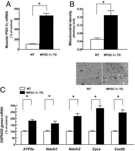
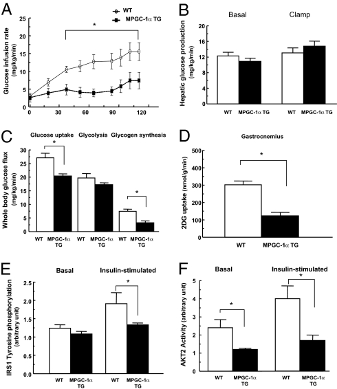
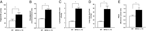
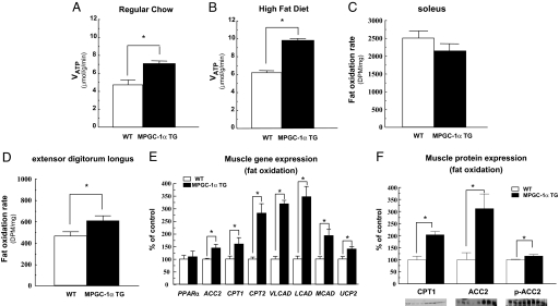
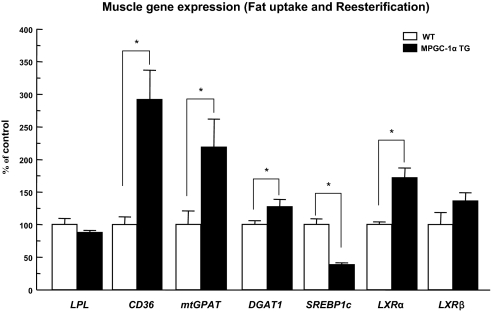
Peroxisome proliferator-activated receptor-gamma coactivator (PGC)-1alpha has been shown to play critical roles in regulating mitochondria biogenesis, respiration, and muscle oxidative phenotype. Furthermore, reductions in the expression of PGC-1alpha in muscle have been implicated in the pathogenesis of type 2 diabetes. To determine the effect of increased muscle-specific PGC-1alpha expression on muscle mitochondrial function and glucose and lipid metabolism in vivo, we examined body composition, energy balance, and liver and muscle insulin sensitivity by hyperinsulinemic-euglycemic clamp studies and muscle energetics by using (31)P magnetic resonance spectroscopy in transgenic mice. Increased expression of PGC-1alpha in muscle resulted in a 2.4-fold increase in mitochondrial density, which was associated with an approximately 60% increase in the unidirectional rate of ATP synthesis. Surprisingly, there was no effect of increased muscle PGC-1alpha expression on whole-body energy expenditure, and PGC-1alpha transgenic mice were more prone to fat-induced insulin resistance because of decreased insulin-stimulated muscle glucose uptake. The reduced insulin-stimulated muscle glucose uptake could most likely be attributed to a relative increase in fatty acid delivery/triglyceride reesterfication, as reflected by increased expression of CD36, acyl-CoA:diacylglycerol acyltransferase1, and mitochondrial acyl-CoA:glycerol-sn-3-phosphate acyltransferase, that may have exceeded mitochondrial fatty acid oxidation, resulting in increased intracellular lipid accumulation and an increase in the membrane to cytosol diacylglycerol content. This, in turn, caused activation of PKC, decreased insulin signaling at the level of insulin receptor substrate-1 (IRS-1) tyrosine phosphorylation, and skeletal muscle insulin resistance.


Conflict of interest statement

The authors declare no conflict of interest.

Figures

Fig. 1.
Fig. 1.
Increased expression of PGC-1α in muscle elevated mitochondrial density and OXPHOS genes. Total RNA was isolated from tibialis anterior muscle of the mice fed a regular diet (n = 5). (A and C) PGC-1α (A) and OXPHOS genes (C) expression was assessed by real-time RT-PCR. (B) Mitochondrial density was counted in EDL muscle (n = 5). (Magnification: ×18,500.) The asterisk indicates mitochondrion. APT5o, ATP synthase; Ndufs, NADH-ubiquinone oxidoreductase; Cycs, cytochrome c; Cox5b, cytochrome c oxidase.
Fig. 2.
Fig. 2.
Increased expression of PGC-1α in skeletal muscle paradoxically decreased peripheral insulin sensitivity in high-fat fed mice. (A–D) Peripheral and hepatic insulin sensitivity was assessed by hyperinsulinemic-euglycemic clamps. (A) Glucose infusion rates. (B) Hepatic glucose production. (C) Whole-body glucose uptake, glycolysis, and glycogen synthesis. (D) skeletal muscle (gastrocnemius) glucose uptake. (E and F) Consistent with decreased muscle insulin sensitivity, IRS1 tyrosine phosphorylation and AKT2 activity in skeletal muscle (gastrocnemius) were decreased in MPGC-1α TG compared with WT mice. IRS1 tyrosine phosphorylation and Akt2 activity were assessed 14 min after i.p. insulin injection of 1 unit/kg body weight. n = 6–8 per group.
Fig. 3.
Fig. 3.
Fat metabolites and membrane translocation of PKCθ were increased in skeletal muscle of MPGC-1α TG mice. (A, C, and D) Triglyceride (A), lysophosphatidic acid (C), and long-chain acyl-CoA (D) levels were significantly increased in muscle (gastrocnemius) in MPGC-1α TG mice. (B and E) Membrane/cytosol ratio of diacylglycerol (B) and PKCθ (E) were significantly higher in muscle (gastrocnemius) of MPGC-1α TG mice compared with WT mice. n = 8 per group.
Fig. 4.
Fig. 4.
Increased expression of PGC-1α in muscle also elevated TRB-3 gene (n = 8) and protein expression (n = 4) in skeletal muscle. Total RNA was isolated from tibialis anterior muscle, TRB-3 gene expression was assessed by real-time RT-PCR (A), and protein expression was confirmed by immunoblot (B).
Fig. 5.
Fig. 5.
In vivo rates of ATP production and ex vivo fat oxidation were increased in the muscle of increased PGC-1α expression. (A and B) Saturation transfer measurements of ATP synthesis rates (VATP) by 31P-MRS in hind limb of mice (n = 6) on regular chow (A) and high-fat diet (B). (C and D) Ex vivo skeletal muscle fat oxidation rate in soleus muscle (C) and EDL muscle (D) from mice (n = 6) fed regular chow. (E) The expression of oxidative/thermogenic genes in tibialis anterior muscle from mice (n = 5–6) fed regular chow assessed by real-time RT-PCR. (F) Immunoblot analysis (n = 4) from selected genes for CPT1, ACC2, and phospho-ACC2 (Ser-219/221). VLCAD, very long-chain acyl-CoA dehydrogenase; LCAD, long-chain acyl-CoA dehydrogenase; MCAD, medium-chain acyl-CoA dehydrogenase; UCP2, uncoupling protein 2.
Fig. 6.
Fig. 6.
The expression of the genes involved in fatty acid uptake and reesterification was increased in tibialis anterior muscle of MPGC-1α TG mice fed regular chow (n = 5–6).

References

    1. Zimmet P, Alberti KG, Shaw J. Global and societal implications of the diabetes epidemic. Nature. 2001;414:782–787. - PubMed
    1. Lillioja S, et al. Insulin resistance and insulin secretory dysfunction as precursors of non-insulin-dependent diabetes mellitus: Prospective studies of Pima Indians. N Engl J Med. 1993;329:1988–1992. - PubMed
    1. DeFronzo RA, Bonadonna RC, Ferrannini E. Pathogenesis of NIDDM: A balanced overview. Diabetes Care. 1992;15:318–368. - PubMed
    1. Boden G, Shulman GI. Free fatty acids in obesity and type 2 diabetes: Defining their role in the development of insulin resistance and β-cell dysfunction. Eur J Clin Invest. 2002;32(Suppl 3):14–23. - PubMed
    1. Kim JK, et al. Tissue-specific overexpression of lipoprotein lipase causes tissue-specific insulin resistance. Proc Natl Acad Sci USA. 2001;98:7522–7527. - PMC - PubMed

Publication types

MeSH terms

LinkOut - more resources