. 2008 Dec 16;105(50):19666-71.
doi: 10.1073/pnas.0807826105. Epub 2008 Dec 9.

Structure of membrane-bound alpha-synuclein from site-directed spin labeling and computational refinement

Affiliations

Structure of membrane-bound alpha-synuclein from site-directed spin labeling and computational refinement

Christine C Jao et al. Proc Natl Acad Sci U S A. .

Abstract

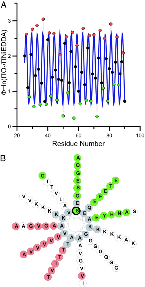
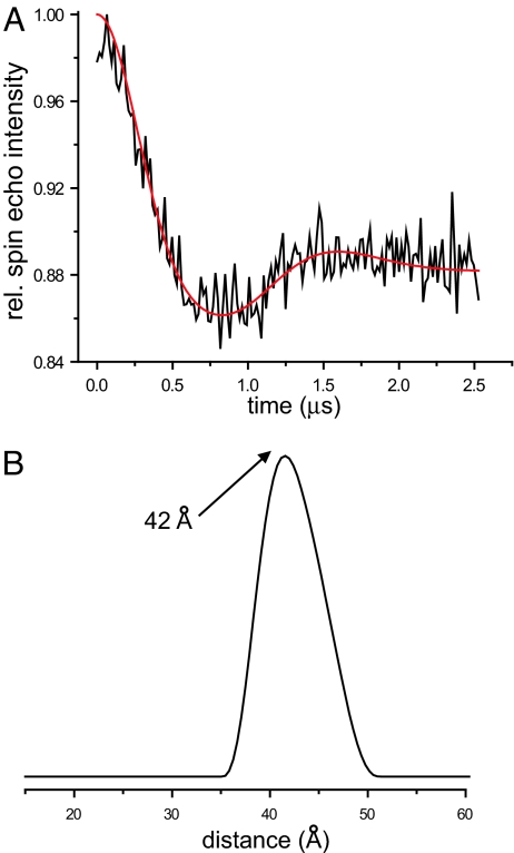
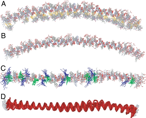
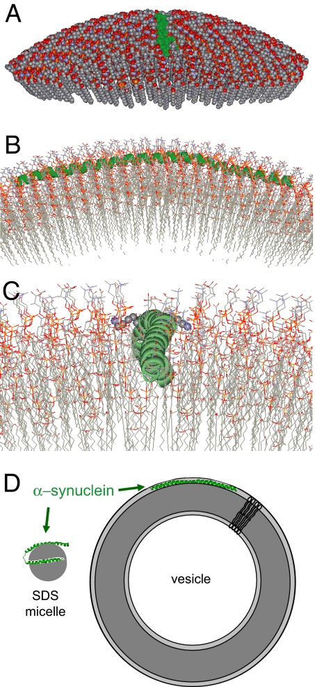
alpha-Synuclein is known to play a causative role in Parkinson disease. Although its physiological functions are not fully understood, alpha-synuclein has been shown to interact with synaptic vesicles and modulate neurotransmitter release. However, the structure of its physiologically relevant membrane-bound state remains unknown. Here we developed a site-directed spin labeling and EPR-based approach for determining the structure of alpha-synuclein bound to a lipid bilayer. Continuous-wave EPR was used to assign local secondary structure and to determine the membrane immersion depth of lipid-exposed residues, whereas pulsed EPR was used to map long-range distances. The structure of alpha-synuclein was built and refined by using simulated annealing molecular dynamics restrained by the immersion depths and distances. We found that alpha-synuclein forms an extended, curved alpha-helical structure that is over 90 aa in length. The monomeric helix has a superhelical twist similar to that of right-handed coiled-coils which, like alpha-synuclein, contain 11-aa repeats, but which are soluble, oligomeric proteins (rmsd = 0.82 A). The alpha-synuclein helix extends parallel to the curved membrane in a manner that allows conserved Lys and Glu residues to interact with the zwitterionic headgroups, while uncharged residues penetrate into the acyl chain region. This structural arrangement is significantly different from that of alpha-synuclein in the presence of the commonly used membrane-mimetic detergent SDS, which induces the formation of two antiparallel helices. Our structural analysis emphasizes the importance of studying membrane protein structure in a bilayer environment.


Conflict of interest statement

The authors declare no conflict of interest.

Figures

Fig. 1.
Fig. 1.
Continuous-wave EPR analysis of singly labeled α-synuclein derivatives indicates the formation of an ordered and continuous helical structure. (A) The ratios of the accessibilities to O2 and NiEDDA for residues 25–90 are summarized by the depth parameter Φ = ln(ΠO2/ΠNiEDDA), with increasing Φ values indicating deeper membrane immersion depth. The blue line indicates the best fit to a cosine function and the resulting periodicity is 3.68 aa per turn (r2 = 0.85), which is close to the theoretically predicted periodicity of 3.67 aa per turn (11 aa per 3 turns = 3.67 aa per turn). (B) The repeat region residues are plotted onto a helical wheel in which 11 aa make up three turns. Lipid-exposed sites (red) fall onto one side, while solvent-exposed sites (green) lie on the opposite side. White circles denote residues with Φ values that are neither maxima nor minima. Residues shown in gray were not tested.
Fig. 2.
Fig. 2.
Intramolecular distances from four-pulse DEER experiments. (A) The baseline corrected time evolution data from a four-pulse DEER experiment for the membrane-bound 56R1/85R1 α-synuclein derivative (black line) were fit by using Tikhonov regularization (34) (red line). The resulting distance distribution is given in B. All results, including those from 16 additional membrane-bound doubly labeled derivatives, are summarized in Table 1 (for data, see Fig. S3).
Fig. 3.
Fig. 3.
Refinement of the membrane-bound structure of α-synuclein by using SAMD with restraints from EPR data. (A) Overlay of nine structures obtained from SAMD calculations using the amino acids 9–89 fragment of α-synuclein, with spin labels added at 26 sites (Fig. S5). In the refined structures, the labels (identifiable by the S–S bond depicted in yellow) are generally positioned on the concave side of the curved protein structure (i.e., oriented into the membrane). The N terminus is on the left of the figure. (B) The refined labeled structures were converted to the respective α-synuclein structures by replacing each label with the normal amino acid in the α-synuclein sequence, with retention of the Cβ position in the label. (C) View of the overlaid structures from above the lipid surface. The 11 lysine residues (blue) are oriented approximately perpendicular to the helical axis, permitting potential interactions with the lipid phosphates. In contrast, the 8 glutamic acid residues (green) are oriented away from the membrane, on the top surface of the α-helix. (D) Ribbon view of the 9 overlaid structures (perspective similar to that in C), indicating the general goodness of fit of the structures and, most importantly, showing the subtle but reproducible superhelical twist in the α-helix that emerged in the SAMD calculations. We speculate that the superhelicity may allow the helix to adopt a 3-11 conformation that permits the helical axis to follow a curved surface over an extended length.
Fig. 4.
Fig. 4.
Representations of the interaction of α-synuclein with a curved lipid surface. (A) Space-filled model of α-synuclein (shown in green) binding to the surface of a lipid vesicle 300 Å in diameter; ≈25% of the outer leaflet of the vesicle is shown. The vesicle was fitted around one of the structures derived from the experimentally restrained SAMD calculations. (B) A closer cross-sectional view of the α-synuclein interaction with the lipid surface, with rotation through 90° from the image in A. The protein (green) follows the curved surface of the vesicle, with the helical axis positioned just below the level of the phosphate groups of the lipids. This position of the protein emerged from the SAMD calculations and is a reflection of the immersion depths obtained from the continuous-wave EPR data. (C) A more detailed image of the protein–lipid interaction, viewed from the same angle as the image in A. The N terminus of the α-helix is in the foreground. Lysine residues 58 and 60 are shown in space-filling format (K58 oriented to the right and K60 to the left of the helix). The image indicates the proximity of the positively charged lysine side chains to the negatively charged phosphate groups. (D) Cartoon representations of the structures of α-synuclein on micelles and SUVs. The small and highly curved micelles cannot accommodate the extended helical structure present on the membrane.
Fig. 5.
Fig. 5.
Comparison of the structure of membrane-bound α-synuclein to that of a right-handed coiled-coil. Overlay of a single helix from tetrabrachion (red, Protein Data Bank ID 1FE6) with that of α-synuclein (green) using Tm-align (41) results in a backbone rmsd of 0.82 Å. The tetrabrachion helices contain 52 aa. Shown are residues 12–62 for α-synuclein (structure 3, which scored highest in the validation (Fig. S6) and residues 2–52 of tetrabrachion. Comparable rmsd values were obtained for all other structures. Overlays with all structures resulted in Tm-scores larger than 0.5, which is an indication of the same fold (41).

References

    1. Cole NB, et al. Lipid droplet binding and oligomerization properties of the Parkinson's disease protein alpha-synuclein. J Biol Chem. 2002;277:6344–6352. - PubMed
    1. Lee HJ, Choi C, Lee SJ. Membrane-bound alpha-synuclein has a high aggregation propensity and the ability to seed the aggregation of the cytosolic form. J Biol Chem. 2002;277:671–678. - PubMed
    1. Lotharius J, Brundin P. Pathogenesis of Parkinson's disease: Dopamine, vesicles and alpha-synuclein. Nat Rev Neurosci. 2002;3:932–942. - PubMed
    1. Zhu M, Li J, Fink AL. The association of alpha-synuclein with membranes affects bilayer structure, stability, and fibril formation. J Biol Chem. 2003;278:40186–40197. - PubMed
    1. Fortin DL, et al. Lipid rafts mediate the synaptic localization of alpha-synuclein. J Neurosci. 2004;24:6715–6723. - PMC - PubMed

Publication types

LinkOut - more resources