

Conformal geometry of the retinal nerve fiber layer
- PMID: 19066221
- PMCID: PMC2604996
- DOI: 10.1073/pnas.0801621105
Conformal geometry of the retinal nerve fiber layer
Abstract
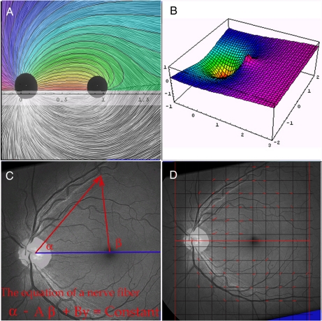
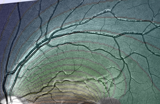
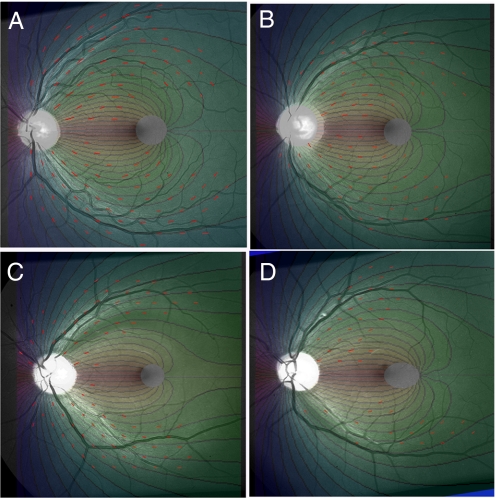
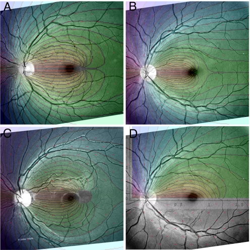
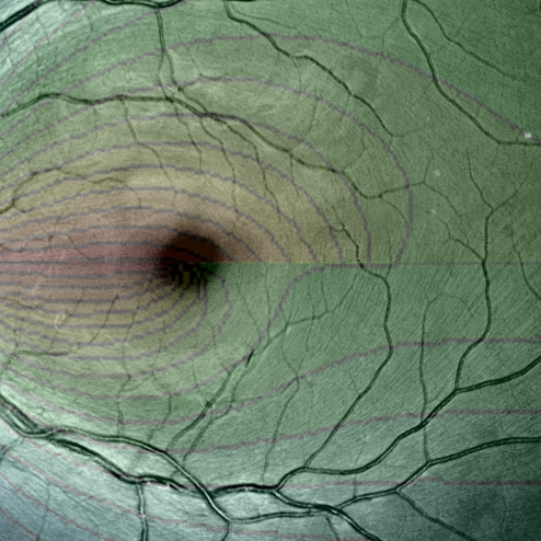
The nerve fiber layer of the human retina is made up of the retinal segments of ganglion cell axons. Its geometry can be described mathematically as a fibration of a 2D domain: a partition of a certain region into smooth curves. Here, we present a simple family of curves that closely models the observed geometry of the nerve fiber layer. For each retina, the pattern depends on 2 parameters, A and B: A computer program determines A and B for a given retina and the theory matches the retina with a standard deviation of approximately 6-8 degrees . These particular curves turn out to be the curves that would be generated if the growing ganglion cell axon tip moved down a gradient toward a source of diffusible neuroattractant at the disk and away from a weaker macular diffusible repellant. Thus, this model provides morphological evidence that diffusible substances provide positional information to the embryonic ganglion cell axons in finding their way to the optic nerve head.
Conflict of interest statement
The authors declare no conflict of interest.
Figures
References
-
- Vogt A. Die Nervenfaserstreifung der menschlichen Netzhaut mit besonderer Berücksichtigung der Differentialdiagnose gegenüber pathologischen streifenförmigen Reflexen (präretinalen Fältelungen) Klin Monatsbl Augenheilkd. 1917;58:399–411.
-
- Behrendt T, Wilson LA. Spectral reflectance photography of the retina. Am J Ophthal. 1965;59:1079–1088. - PubMed
-
- Airaksinen PJ, Nieminen H, Mustonen E. Retinal nerve fiber layer photography with a wide angle fundus camera. Acta Ophthal. 1982;60:362–368. - PubMed
-
- Crick F. Diffusion in embryogenesis. Nature. 1970;225:420–422. - PubMed
-
- Ramon y, Cajal S. La rétine des vertébrés. 1892;9:119. Available at http://web2.bium.univ-paris5.fr/livanc/?cote=ryc007&do=chapitre.
MeSH terms
LinkOut - more resources
Full Text Sources
