. 2009 Mar;17(3):463-71.
doi: 10.1038/mt.2008.269. Epub 2008 Dec 16.

High-efficiency transduction of the mouse retina by tyrosine-mutant AAV serotype vectors

Affiliations

High-efficiency transduction of the mouse retina by tyrosine-mutant AAV serotype vectors

Hilda Petrs-Silva et al. Mol Ther. 2009 Mar.

Abstract

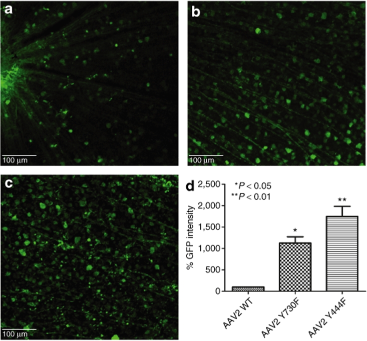
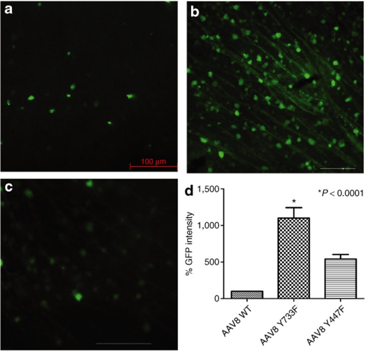
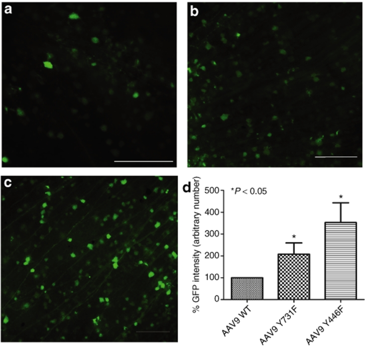
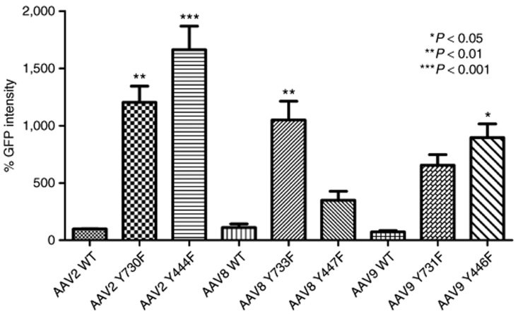
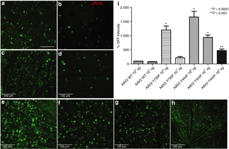
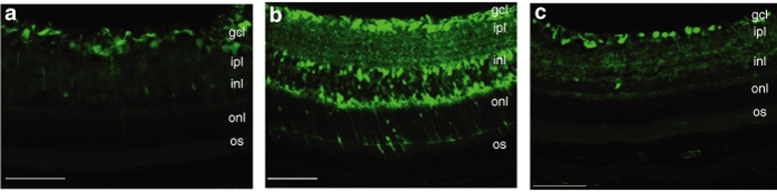
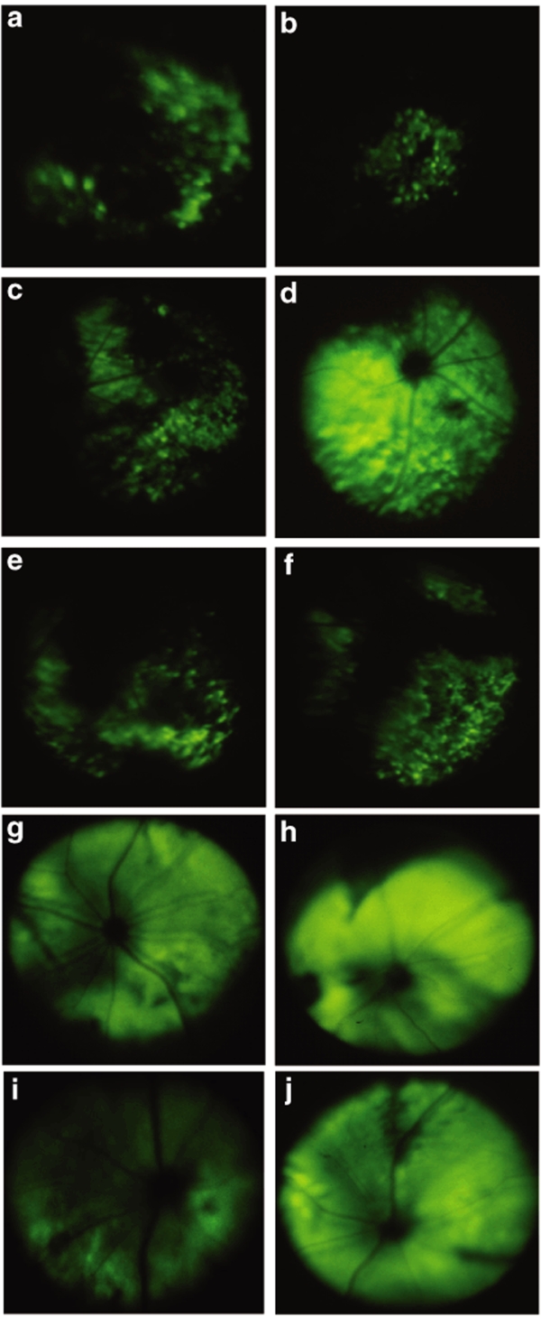
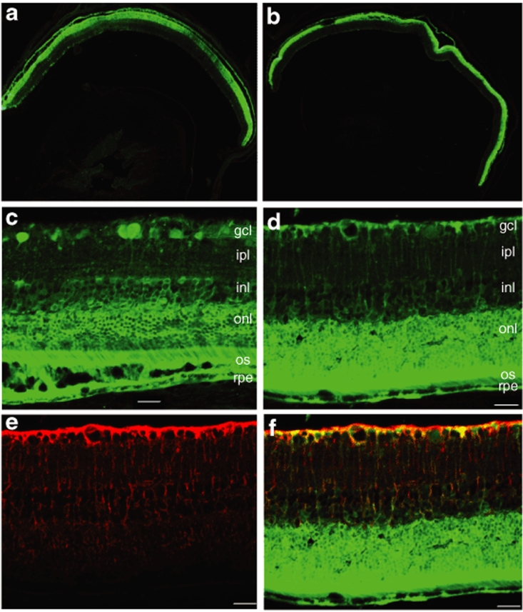
Vectors derived from adeno-associated viruses (AAVs) have become important gene delivery tools for the treatment of many inherited ocular diseases in well-characterized animal models. Previous studies have determined that the viral capsid plays an essential role in the cellular tropism and efficiency of transgene expression. Recently, it was shown that phosphorylation of surface-exposed tyrosine residues from AAV2 capsid targets the viral particles for ubiquitination and proteasome- mediated degradation, and mutations of these tyrosine residues lead to highly efficient vector transduction in vitro and in vivo. Because the tyrosine residues are highly conserved in other AAV serotypes, in this study we evaluated the intraocular transduction characteristics of vectors containing point mutations in surface- exposed capsid tyrosine residues in AAV serotypes 2, 8, and 9. Several of these novel AAV mutants were found to display a strong and widespread transgene expression in many retinal cells after subretinal or intravitreal delivery compared with their wild-type counterparts. For the first time, we show efficient transduction of the ganglion cell layer by AAV serotype 8 or 9 mutant vectors, thus providing additional tools besides AAV2 for targeting these cells. These enhanced AAV vectors have a great potential for future therapeutic applications for retinal degenerations and ocular neovascular diseases.


Figures

<b>Figure 1</b>
Figure 1
Analysis of enhanced green fluorescent protein (EGFP) expression 2 weeks after intravitreal delivery of equal doses of wild-type (WT) scAAV2-CBA-EGFP or its tyrosine mutants. (a–c) Immunohistochemistry for EGFP in flat-mount retinas infected with (a) WT AAV2 vector, (b) mutant Y730F, or (c) mutant Y444F. Calibration bar 100 µm. All pictures were taken with the same exposure time to evaluate EGFP intensity using ImageJ. (d) Values indicate percentage of EGFP intensity of the mutants compared with WT. Both type 2 tyrosine mutants showed a statistically significant increase in EGFP intensity with *P < 0.05 for Y730F; **P < 0.01 for Y444F versus WT AAV2. Statistical analyses were performed with one-way ANOVA plus Dunnett's multiple-range test compared to the control group (WT scAAV2). CBA, chicken β-actin; scAAV, self-complementary adeno-associated virus.
<b>Figure 2</b>
Figure 2
Analysis of enhanced green fluorescent protein (EGFP) expression 2 weeks after intravitreal delivery of equal doses of wild-type (WT) scAAV8-CBA-EGFP or its tyrosine mutants. (a–c) Immunohistochemistry for EGFP in flat-mount retinas infected with WT (a) AAV8 vector, (b) mutant Y733F, or (c) mutant Y447F. Calibration bar 100 µm. All pictures were taken with the same exposure time to evaluate EGFP intensity with ImageJ. (d) Values indicate percentage of EGFP intensity of the mutants compared with WT. Only tyrosine-mutant Y733F showed a statistically significant elevation in EGFP intensity (*P < 0.0001) compared with WT AAV8. Statistical analyses were performed with one-way ANOVA plus Dunnett's multiple-range test compared to the control group (WT scAAV8). CBA, chicken β-actin; scAAV, self- complementary adeno-associated virus.
<b>Figure 3</b>
Figure 3
Analysis of enhanced green fluorescent protein (EGFP) expression 2 weeks after equal doses of intravitreal delivery of wild-type self-complementary adeno-associated virus 9 (WT scAAV9) vector or its tyrosine mutants. (a–c) Immunohistochemistry for EGFP in flat-mount retinas infected with (a) WT AAV9 vector, (b) mutant Y731F, or (c) mutant Y446F. Calibration bar 100 µm. All pictures were taken with the same exposure time to evaluate EGFP intensity with ImageJ. (d) Values indicate percentage of EGFP intensity of the mutants compared with WT. Both tyrosine mutants of type 9 showed statistically significant increase in EGFP intensity with *P < 0.0001 for both mutants versus WT AAV9. Statistical analyses were performed with one-way ANOVA plus Dunnett's multiple-range test compared to the control group (WT scAAV9).
<b>Figure 4</b>
Figure 4
Comparison of enhanced green fluorescent protein (EGFP) intensity of all serotypes and their mutants with wild-type self-complementary adeno-associated virus 2 (WT scAAV2) vector 2 weeks after intravitreal delivery. Retinal flat mounts were immunolabeled with polyclonal EGFP antibody. Pictures were taken with the same exposure time to evaluate the intensity, with ImageJ. Values indicate percentage of EGFP intensity compared with wild-type AAV2 vector. Type 2 Y444F showed the highest GFP expression enhancement (***P < 0.001); type 2 mutant Y730F and type 8 Y733F also significantly EGFP expression (**P < 0.01); type 9 mutant Y446F also had a higher level of EGFP (*P < 0.05). Statistical analyses were performed with one-way ANOVA plus Dunnett's multiple-range test compared to the control group (WT scAAV2).
<b>Figure 5</b>
Figure 5
Enhanced green fluorescent protein (EGFP) expression in murine retinas intravitreally injected with serial dilutions of the two strongest mutants compared with wild type (WT). Retina flat-mount immunostained for EGFP 2 weeks after intravitreal delivery of WT-2 at 109 vector genomes (vg) (a) and 107 vg (b); mutant Y730F at 109 vg (c) and 107 vg (d); and mutant Y444F at 109 vg (e), 107 vg (f), and 105 vg at higher magnification (g) and lower magnification (h), providing an assessment of the extent (area) of transduction. Calibration bar 100 µm. EGFP intensity was evaluated with ImageJ and values indicate percentage of EGFP intensity compared with WT adeno-associated virus 2 (AAV2) vector at the standard concentration of 109 vg (i). Statistical analyses were performed with one-way ANOVA plus Dunnett's multiple-range test compared to the control group (WT scAAV2).
<b>Figure 6</b>
Figure 6
Fluorescence microscopic evaluation of enhanced green fluorescent protein (EGFP) expression in transverse sections of retinal tissue 2 weeks after intravitreal injection. Immunostaining for EGFP in sections of the retina after delivery of (a) wild-type self-complementary adeno-associated virus 2 (WT scAAV2), (b) serotype 2 tyrosine-mutant Y444F, and (c) serotype 2 tyrosine-mutant Y730F. Note intense EGFP staining throughout all retinal layers with Y444F mutant and predominant EGFP staining in the GCL with WT-2 and Y730F. Calibration bar 100 µm. gcl, ganglion cell layer; ipl, inner plexiform layer; inl, inner nuclear layer; onl, outer nuclear; os, outer segment.
<b>Figure 7</b>
Figure 7
Fundus photos depicting in vivo enhanced green fluorescent protein (EGFP) fluorescence in adult mouse retinas after subretinal delivery of scAAV-CBA-EGFP viral vectors. At 10 days postinjection, fluorescence was the strongest and most widespread for serotype 8 Y733F (d), and it was only seen in patchy areas of the fundus for serotype 2 Y444F (a) and Y730F (b), wild-type 8 (WT-8) (c), WT-9 (e), and serotype 9 Y446F (f). Representative fundus photographs at 17 days postinjection are shown for WT-8 (g), serotype 8 Y733F (h), serotype 2 Y444F (i), and serotype 9 Y446F (j). CBA, chicken β-actin; scAAV, self-complementary adeno-associated virus.
<b>Figure 8</b>
Figure 8
Analysis of enhanced green fluorescent protein (EGFP) expression in frozen retinal sections by immunohistochemistry at 1 month following subretinal injections with highly efficient Tyr/Phe mutant adeno-associated virus vectors. Representative sections depicting widespread and intense EGFP fluorescence throughout the retina after transduction with (a) serotype 2 Y444F or (b) serotype 8 Y733F. The images are oriented with the vitreous toward the bottom and the photoreceptor layer toward the top. Calibration bar 500 µm. (c) EGFP fluorescence in photoreceptors, retinal pigment epithelial (RPE), and ganglion cells from mouse eyes injected subretinally with serotype 2 Y444F; (d) EGFP fluorescence in photoreceptors, RPE, and Müller cells after serotype 8 Y733F delivery. (e) Detection of Müller cells processes (red) by immunostaining with a glutamine synthetase (GS) antibody. (f) Merged image showing colocalization of EGFP fluorescence (green) and GS staining (red) in retinal sections from eyes treated with serotype 8 Y733F. Calibration bar 100 µm. gcl, ganglion cell layer; ipl, inner plexiform layer; inl, inner nuclear layer; onl, outer nuclear; os, outer segment.

References

    1. Goncalves MA. Adeno-associated virus: from defective virus to effective vector. Virol J. 2005;2:43. - PMC - PubMed
    1. Xie Q, Bu W, Bhatia S, Hare J, Somasundaram T, Azzi A, et al. The atomic structure of adeno-associated virus (AAV-2), a vector for human gene therapy. Proc Natl Acad Sci USA. 2002;99:10405–10410. - PMC - PubMed
    1. Gao G, Vandenberghe LH, Alvira MR, Lu Y, Calcedo R, Zhou X, et al. Clades of adeno-associated viruses are widely disseminated in human tissues. J Virol. 2004;78:6381–6388. - PMC - PubMed
    1. Schmidt M, Voutetakis A, Afione S, Zheng C, Mandikian D., and , Chiorini JA. Adeno-associated virus type 12 (AAV12): a novel AAV serotype with sialic acid- and heparan sulfate proteoglycan-independent transduction activity. J Virol. 2008;82:1399–1406. - PMC - PubMed
    1. Wu Z, Asokan A., and , Samulski RJ. Adeno-associated virus serotypes: vector toolkit for human gene therapy. Mol Ther. 2006;14:316–327. - PubMed

Publication types

LinkOut - more resources