. 2009 Feb;17(2):269-77.
doi: 10.1038/mt.2008.266. Epub 2008 Dec 9.

Targeting hypoxia-inducible factor-1alpha with Tf-PEI-shRNA complex via transferrin receptor-mediated endocytosis inhibits melanoma growth

Affiliations

Targeting hypoxia-inducible factor-1alpha with Tf-PEI-shRNA complex via transferrin receptor-mediated endocytosis inhibits melanoma growth

Yeqiang Liu et al. Mol Ther. 2009 Feb.

Abstract

Malignant melanoma (MM) is a major public health problem. The development of effective, systemic therapies for MM is highly desired. We showed here that the transferrin receptor (TfR) was a suitable surface marker for targeting of gene therapy in MM and that the hypoxia-inducible factor-1alpha (HIF-1alpha) was an attractive therapeutic molecular target in MM. We observed that inhibition of HIF-1alpha blocked cell proliferation and induced cell apoptosis in vitro. We then showed that a transferrin-polyethylenimine-HIF-1alpha-short-hairpin RNA (Tf-PEI-HIF-1alpha-shRNA) complex could target MM specifically and efficiently both in vivo and in vitro, exploiting the high expression of the TfR in MM. The systemic delivery of sequence-specific small-interfering RNA (siRNA) against HIF-1alpha by the Tf- PEI-HIF-1alpha-shRNA complex dramatically inhibited tumor growth in the A375 MM xenograft model. The underlying concept of transfecting a HIF-1alpha shRNA expression vector complexed with Tf-PEI to block HIF-1alpha holds promise as a clinical approach to gene therapy for MM.


Figures
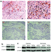
<b>Figure 1</b>
Figure 1
Hypoxia-inducible factor-1α (HIF-1α) expression in malignant melanoma (MM). (a) HIF-1α staining in MM tissues (A1 and A2) and normal nevus (A3 and A4). A strong immune reaction in high-grade MM and a negative immune reaction in normal nevus were typically observed by immunohistochemistry (A1 and A3 ×100 magnifications, A2 and A4 ×200 magnifications). (b) Western blot analysis of HIF-1α expression in the tissue of MM (T) and normal nevus (N). MM tissues had more abundant expression of HIF-1α than did normal nevus. (c) Western blot analysis of HIF-1α expression in A375, A875, and KZ28 cells under hypoxia and normoxia. More abundant expression of HIF-1α protein was observed in A375, A875, and KZ28 cells under hypoxic conditions than under normoxic conditions. Results showed a typical experiment from three performed.
<b>Figure 2</b>
Figure 2
Hypoxia-inducible factor-1α (HIF-1α) short-hairpin RNAs (shRNAs) silencing of HIF-1α in malignant melanoma (MM) cell lines in vitro and its effects. (a) HIF-1α shRNAs downregulated HIF-1α expression in A375 cells. Comparison of HIF-1α messenger RNA (mRNA) levels of different HIF-1α shRNA-treated A375 cells revealed by real-time PCR at different time points. The expression level of HIF-1α was compared to the level of gene expression found in nontransfected negative controls, arbitrarily assigned the value 1. Bars represent the fold reduction in gene expression over the expression level in the nontransfected A375 cells, and shRNA1, shRNA2, and shRNA3 decreased HIF-1α mRNA by 74, 10, and 32%, respectively, in the A375 cells in the 24 hours after transfection. (b) The effects of shRNA1-mediated silencing of HIF-1α gene expression on the proliferation of A375 cells. Viable cells were determined by the MTT assay. The shRNA1-A375 stable clone, the negative control–scrambled shRNA-A375 stable clone, and untreated A375 cells (5 × 10³) were plated on 96-well plates and evaluated by the MTT assay at 48 hours. Each bar represents the mean value of three identical wells from an experiment representative of three trials with different cell cultures. For all experiments combined, the shRNA1-A375 stable clone was significantly less than for the negative control–scrambled shRNA-A375 stable clone and untreated A375 cells. (c) The effects of shRNA1-mediated silencing of HIF-1α gene expression on the apoptosis of A375 cells. Cells in the bottom left quadrant represent viable cells (low annexin V-Alexa and BoBo-1 staining); cells in the bottom right quadrant represent early apoptotic cells (high annexin V-Alexa staining but low BoBo-1 staining); and cells in the top right quadrant represent late apoptotic cells (high annexin V-Alexa and BoBo-1 staining). The percentage of cells in each quadrant is indicated in the right up quadrant of the panels. Shown are representative data from one of three independent experiments with samples in triplicate. Increases in the percentage of apoptotic cells in the shRNA1-A375 stable clone compared with the negative control–scrambled shRNA-A375 stable clone and untreated A375 cells were of statistical significance (P = 0.003).
<b>Figure 3</b>
Figure 3
Transferrin receptor (TfR) expression in malignant melanoma (MM) tissues and cell lines. (a) TfR staining in the tissues of MM (A1 and A2) and normal nevus (A3 and A4). A strong immune reaction in MM and a weak immune reaction in normal nevus were typically observed by immunohistochemistry (A1 and A3 ×100 magnification; A2 and A4 ×200 magnification). (b) TfR expression in the MM cell lines A375, A875, and KZ28 and in the human ovarian cancer cell line A2780 by flow cytometry. The levels of the cell surface TfR in A375, A875, and KZ28 cells are shown in the right top (B1: A2780, B2: A375, B3: A875, B4: KZ28).
<b>Figure 4</b>
Figure 4
Analysis of tumor-targeted distribution of the Tf–PEI–shRNA complex after administration. (a) Tumor-targeted distribution in vitro. Approximately ~50% of the A375 cells took up Tf–PEI–shRNA, whereas the A2780 cells did not. There was no difference in uptake between the A2780 and A375 cells when short-hairpin RNA (shRNA) was transfected by Lipofectamine2000. Neither the A2780 nor A375 cells appreciably took up shRNA-GFP by themselves or when mixed with transferring (Tf) lacking polyethylenimine (PEI) or with unmodified PEI. (b) Tumor-targeted distribution in vivo. Green fluorescent protein (GFP) messenger RNA (mRNA) expression in tumor tissues and major organs of nude mice bearing A375 or A2780 tumors and injected intravenous with Tf–PEI–shRNA was determined by the SYBR Green real-time RT-PCR assay. The method of quantitation of GFP mRNA was described in Materials and Methods. (b1) GFP mRNA distribution in tissues and major organs of A375 tumor–bearing nude mice. (b2) GFP mRNA distribution in tissues and major organs of A2780 tumor–bearing nude mice. The highest amounts of GFP mRNA were detected in tumors and only small amounts of GFP mRNA were detected in major organs such as liver, lungs, heart, and kidneys in the A375 tumor–bearing mice at a time point of 24 hours after the single injection (P = 0.004). The GFP mRNA distribution in tumor tissues and major organs of A2780 tumor–bearing mice revealed no significant differences (P = 0.496).
<b>Figure 5</b>
Figure 5
Observation of the growth rate of malignant melanoma (MM) xenograft tumors after injection with Tf–PEI–shRNA1 in vivo. Each point represents the mean volume ± SD. N = 12 for the shRNA1 and scrambled shRNA–injected groups (n = 12) and n = 6 for the untreated group. One arrow represents the day of the Tf–PEI–shRNA injection, and two arrows is the time of killing of the mice (**P < 0.001). (a) The therapeutic potential of the Tf–PEI–shRNA1 complex in the A375 tumor xenograft. At the time point of killing, for the A375 group, the tumors in the control second and third groups had volumes of 610 ± 145 mm3 and 655 ± 90 mm3, respectively, which were 7.5-fold larger than the starting volume, whereas the A375 tumors of the mice injected with the Tf–PEI–shRNA1 complex had a volume of 310 ± 90 mm3, resulting in a volume increase of only 3.9-fold. The tumor growth delay was statistically significant (P = 0.0014) from day 5 after the beginning of therapy until the day the mice were killed. (b) The effect of the Tf–PEI–shRNA1 complex in the A2780 tumor xenograft. At the time point of sacrificing, the tumors for the A2780 xenografts in the three groups had volumes of 710 ± 145, 755 ± 90, and 695 ± 120 mm3, respectively, which were 7.5-fold larger than the starting volume. A tumor growth delay was not found in the therapeutic group (P = 0.2460). Tf, transferring; PEI, polyethylenimine; shRNA, short-hairpin RNA.
<b>Figure 6</b>
Figure 6
Detection of hypoxia-inducible factor-1α (HIF-1α) knockdown in tumor sections after systemic Tf–PEI–HIF-1α–shRNA complex administration. (a) HIF-1α expression was determined by immunohistochemistry. HIF-1α expression in the Tf–PEI–shRNA1 complex group was significantly lower than in the Tf–PEI–HIF-1α-scrambled shRNA and control groups in the nude mice bearing A375 s.c.xenograft tumors. No significant differences in HIF-1α expression were found between the three groups in the nude mice bearing A2780 s.c.xenograft tumors (×200 magnification). (b) HIF-1α expression determined by western blotting. The results as determined by western blotting were in agreement with those determined by immunohistochemistry. Tf, transferring; PEI, polyethylenimine; shRNA, short-hairpin RNA.

References

    1. Scala S, Ierano C, Ottaiano A, Franco R, La Mura A, Liguori G, et al. CXC chemokine receptor 4 is expressed in uveal malignant melanoma and correlates with the epithelioid-mixed cell type. Cancer Immunol Immunother. 2007;56:1589–1595. - PMC - PubMed
    1. Markovic SN, Erickson LA, Rao RD, Weenig RH, Pockaj BA, Bardia A, et al. Malignant melanoma in the 21st century, part 1: epidemiology, risk factors, screening, prevention, and diagnosis. Mayo Clin Proc. 2007;82:364–380. - PubMed
    1. Becker JC, Kirkwood JM, Agarwala SS, Dummer R, Schrama D., and , Hauschild A. Molecularly targeted therapy for melanoma: current reality and future options. Cancer. 2006;107:2317–2327. - PubMed
    1. Tao J, Tu YT, Huang CZ, Feng AP, Wu Q, Lian YJ, et al. Inhibiting the growth of malignant melanoma by blocking the expression of vascular endothelial growth factor using an RNA interference approach. Br J Dermatol. 2005;153:715–724. - PubMed
    1. Kamat CD, Green DE, Curilla S, Warnke L, Hamilton JW, Sturup S, et al. Role of HIF signaling on tumorigenesis in response to chronic low-dose arsenic administration. Toxicol Sci. 2005;86:248–257. - PubMed

Publication types

MeSH terms