. 2009 Feb;17(2):253-61.
doi: 10.1038/mt.2008.264. Epub 2008 Dec 9.

Cardiac expression of a mini-dystrophin that normalizes skeletal muscle force only partially restores heart function in aged Mdx mice

Affiliations

Cardiac expression of a mini-dystrophin that normalizes skeletal muscle force only partially restores heart function in aged Mdx mice

Brian Bostick et al. Mol Ther. 2009 Feb.

Abstract

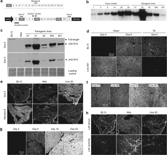
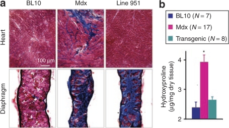
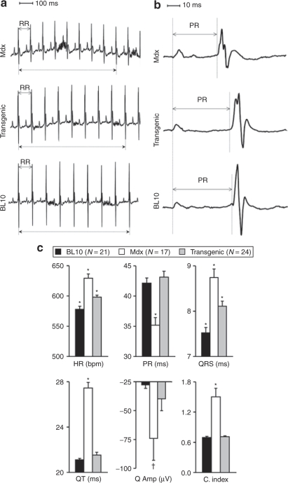
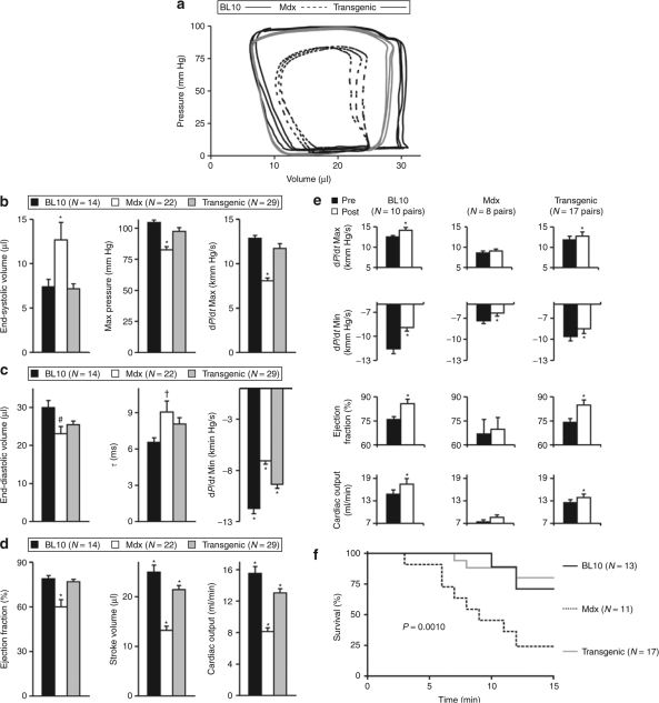
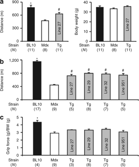
Duchenne muscular dystrophy (DMD) affects both skeletal and cardiac muscle. It is currently unclear whether the strategies developed for skeletal muscle can ameliorate cardiomyopathy. Synthetic mini-/micro-dystrophin genes have yielded impressive skeletal muscle protection in animal models. The 6-kb DeltaH2-R19 minigene is particularly promising because it completely restores skeletal muscle force to wild-type levels. Here, we examined whether expressing this minigene in the heart, but not skeletal muscle, could normalize cardiac function in the mdx model of DMD cardiomyopathy. Transgenic mdx mice were generated to express the DeltaH2-R19 minigene under the control of the alpha-myosin heavy-chain promoter. Heart structure and function were examined in adult and very old mice. The DeltaH2-R19 minigene enhanced cardiomyocyte sarcolemmal strength and prevented myocardial fibrosis. It also restored the dobutamine response and enhanced treadmill performance. Surprisingly, heart-restricted DeltaH2-R19 minigene expression did not completely normalize electrocardiogram and hemodynamic abnormalities. Overall, systolic function and ejection fraction were restored to normal levels but stroke volume and cardiac output remained suboptimal. Our results demonstrate that the skeletal muscle-proven DeltaH2-R19 minigene can correct cardiac histopathology but cannot fully normalize heart function. Novel strategies must be developed to completely restore heart function in DMD.


Figures

<b>Figure 1</b>
Figure 1
Characterization of cardiac-specific ΔH2-R19 mini-dystrophin transgenic mice. (a) Schematic outline of the transgenic construct. Dotted lines denote the regions deleted (from H2 to R19) from the full-length gene. The locations of the PCR primers (DL254 and DL255) and the origin of the Southern probe are marked. The regions recognized by Dys-2, Dys-3, and Mandys-8 antibodies are also marked. Asterisk, Dys-3 antibody only recognizes human dystrophin. C, C-terminal domain; CR, cysteine-rich domain; H, hinge; N, N-terminal domain; numerical numbers, spectrin-like repeats. (b) A representative Southern blot result from transgenic lines 31, 32, 33, 950, and 951. (c) A representative heart western blot from BL10, mdx, and transgenic lines 27, 31, 32, 950, and 951. Top panel, Dys-2 antibody reveals both the endogenous full-length mouse dystrophin in the BL10 heart and human mini-dystrophin in transgenic mice; middle panel, Dys-3 antibody only reveals human mini-dystrophin; bottom panel, loading control (rapid blue staining of a duplicate gel). (d) Representative Dys-2 and Dys-3 immunofluorescence staining images of the hearts and the tibialis anterior (TA) muscles from BL10 and transgenic line 951 mice. (e) Representative Dys-3 and Mandys-8 immunofluorescence staining images of the left atrium from BL10, mdx, and transgenic line 30 mice. (f) Representative Dys-3 immunofluorescence staining photomicrographs from transgenic lines 27, 30, 31, and 951. (g) Representative photomicrographs of Dys-2 heart immunofluorescence staining from line 32 at different postnatal development stages. (h) Representative left-heart utrophin immunofluorescence staining images from BL10, mdx, and transgenic line 30 mice. To facilitate the comparison of relative expression levels, images in the same panel were taken under the exact same exposure conditions.
<b>Figure 2</b>
Figure 2
ΔH2-R19 mini-dystrophin stabilizes sarcolemmal integrity in the heart. (a) Representative heart Evans blue dye (EBD) uptake photomicrographs. Numbers denote transgenic line. Neg, transgene-negative littermate from the same breeding. (b) Quantification of the EBD-positive area. N, sample size. Asterisk, results from mdx mice and transgene-negative littermates are significantly different from BL10 and transgenic mice. There was no statistical difference between BL10 and different lines of transgenic mice.
<b>Figure 3</b>
Figure 3
Cardiac-specific ΔH2-R19 mini-dystrophin expression eliminates fibrosis in the heart but not in the diaphragm. (a) Representative Masson trichrome staining photomicrographs from the heart (top row) and the diaphragm (bottom row) in 22-month-old BL10, mdx, and transgenic line 951 mice. Connective tissue is in blue color. (b) Heart hydroxyproline content normalized by dry tissue weight. N, sample size. Asterisk, result from mdx mice was significantly different from both BL10 and transgenic mice. There was no statistical difference between BL10 and transgenic mice.
<b>Figure 4</b>
Figure 4
Electrocardiogram (ECG) profiles in 22-month-old BL10, mdx, and transgenic mice. (a) Representative serial lead II ECG tracings. The dotted lines mark the time from the first to ninth R waves in each ECG tracing. (b) Representative serial lead II ECG tracings. (c) Quantitative evaluation of ECG profile. The results for transgenic mice are combined data from line 27 (five mice), line 29 (four mice), line 32 (eight mice), line 30 (one mouse), and line 951 (six mice). C. index, the cardiomyopathy index; HR, heart rate; Q Amp, the Q wave amplitude. Asterisk, significantly different from the other two groups; dagger, mdx was significantly different from BL10.
<b>Figure 5</b>
Figure 5
Characterization of hemodynamic changes in 22-month-old male BL10, mdx, and transgenic mice. (a) Representative PV loops. (b) Baseline hemodynamic profile during systole. (c) Baseline diastolic profile. (d) Overall heart function evaluation. The transgenic group in panels b to d includes four mice from line 27, eight mice from line 29, one mouse from line 30, one mouse from line 31, six mice from line 32, two mice from line 950, and seven mice from line 951. (e) Hemodynamic responses to dobutamine challenge. Pre, hemodynamic information before dobutamine administration; post, hemodynamic information at 5 minutes after dobutamine administration. The transgenic group in panels e and f includes six mice from line 27, five mice from line 29, one mouse from line 30, one mouse from line 31, and four mice from line 951. Max, maximal; Min, minimal. Asterisk (a–c), significantly different from the other two groups; asterisk (e), significantly different from pre-dobutamine stress; hash, significantly different from BL10 on ANOVA analysis; dagger, multiple group comparison by ANOVA shows no significant difference among three groups. However, t-test shows a significant difference between BL10 and mdx mice. (f) Kaplan–Meier survival analysis in mice stressed with dobutamine.
<b>Figure 6</b>
Figure 6
Cardiac-specific ΔH2-R19 mini-dystrophin expression improves uphill running performance without enhancing limb muscle strength. (a) Running distance and body weight in 8-month-old male mice. (b) Running distance in 18-month-old male mice (note, different treadmill protocols were used for 8- and 18-month-old mice). (c) Body weight normalized forelimb grip-strength in 18-month-old mice. N, sample size; Tg, transgenic mice; asterisk, significantly higher than all other groups; hash, significantly higher than that of mdx mice but lower than that of BL10 mice.

References

    1. American Academy of Pediatrics Section on Cardiology and Cardiac Surgery (2005) Cardiovascular health supervision for individuals affected by Duchenne or Becker muscular dystrophy. Pediatrics. 116:1569–1573. - PubMed
    1. Baxter P. Treatment of the heart in Duchenne muscular dystrophy. Dev Med Child Neurol. 2006;48:163. - PubMed
    1. Duan D.Challenges and opportunities in dystrophin-deficient cardiomyopathy gene therapy Hum Mol Genet 200615R253–R261.spec. 2 - PMC - PubMed
    1. Ervasti JM. Dystrophin, its interactions with other proteins, and implications for muscular dystrophy. Biochim Biophys Acta. 2007;1772:108–117. - PubMed
    1. Warner LE, DelloRusso C, Crawford RW, Rybakova IN, Patel JR, Ervasti JM, et al. Expression of Dp260 in muscle tethers the actin cytoskeleton to the dystrophin-glycoprotein complex and partially prevents dystrophy. Hum Mol Genet. 2002;11:1095–1105. - PubMed

Publication types

LinkOut - more resources