

The effects of GPX-1 knockout on membrane transport and intracellular homeostasis in the lens
- PMID: 19067024
- PMCID: PMC4470390
- DOI: 10.1007/s00232-008-9141-5
The effects of GPX-1 knockout on membrane transport and intracellular homeostasis in the lens
Abstract
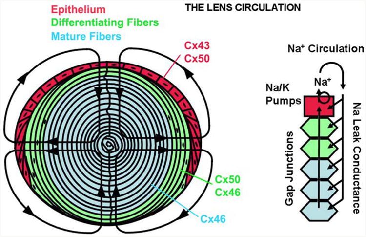
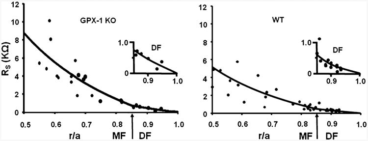
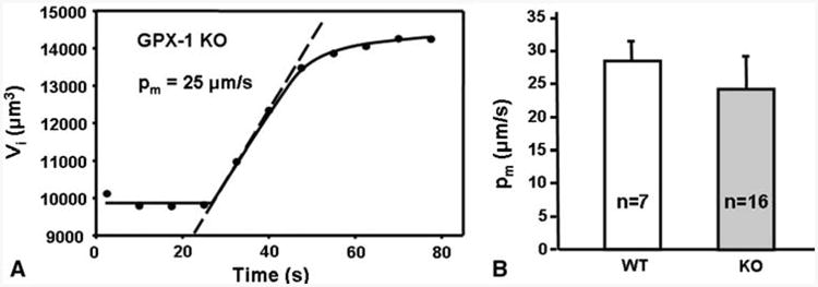
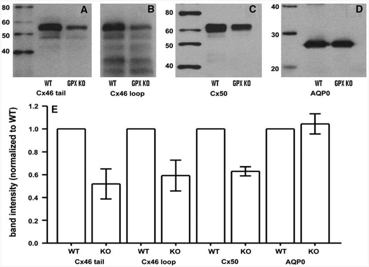
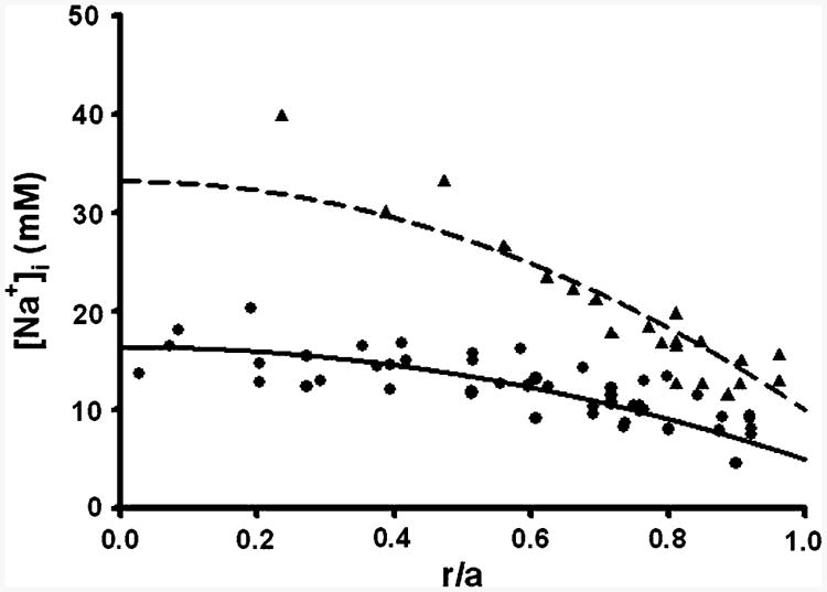
Glutathione peroxidase-1 (GPX-1) is an enzyme that protects the lens against H2O2-mediated oxidative damage. The purpose of the present study was to determine the effects of GPX-1 knockout (KO) on lens transport and intracellular homeostasis. To investigate these lenses we used (1) whole lens impedance studies to measure membrane conductance, resting voltage and fiber cell gap junction coupling conductance; (2) osmotic swelling of fiber cell membrane vesicles to determine water permeability; and (3) injection of Fura2 and Na+-binding benzofuran isophthalate (SBFI) into fiber cells to measure [Ca2+]i and [Na+]i, respectively, in intact lenses. These approaches were used to compare wild-type (WT) and GPX-1 KO lenses from mice around 2 months of age. There were no significant differences in clarity, size, resting voltage, membrane conductance or fiber cell membrane water permeability between WT and GPX-1 KO lenses. However, in GPX-1 KO lenses, coupling conductance was 72% of normal in the outer shell of differentiating fibers and 45% of normal in the inner core of mature fibers. Quantitative Western blots showed that GPX-1 KO lenses had about 50% as much labeled Cx46 and Cx50 protein as WT, whereas they had equivalent labeled AQP0 protein as WT. Both Ca2+ and Na+ accumulated significantly in the core of GPX-1 KO lenses. In summary, the major effect on lens transport of GPX-1 KO was a reduction in gap junction coupling conductance. This reduction affected the lens normal circulation by causing [Na+]i and [Ca2+]i to increase, which could increase cataract susceptibility in GPX-1 KO lenses.
Figures


Similar articles
-
The effects of age on lens transport.Invest Ophthalmol Vis Sci. 2013 Nov 1;54(12):7174-87. doi: 10.1167/iovs.13-12593. Invest Ophthalmol Vis Sci. 2013. PMID: 24065810 Free PMC article.
-
Lens gap junctional coupling is modulated by connexin identity and the locus of gene expression.Invest Ophthalmol Vis Sci. 2004 Oct;45(10):3629-37. doi: 10.1167/iovs.04-0445. Invest Ophthalmol Vis Sci. 2004. PMID: 15452070
-
PKCγ, role in lens differentiation and gap junction coupling.Curr Eye Res. 2011 Jul;36(7):620-31. doi: 10.3109/02713683.2011.573899. Epub 2011 May 20. Curr Eye Res. 2011. PMID: 21599470
-
Lens gap junctions in growth, differentiation, and homeostasis.Physiol Rev. 2010 Jan;90(1):179-206. doi: 10.1152/physrev.00034.2009. Physiol Rev. 2010. PMID: 20086076 Free PMC article. Review.
-
Regulation of Connexin Gap Junctions and Hemichannels by Calcium and Calcium Binding Protein Calmodulin.Int J Mol Sci. 2020 Nov 2;21(21):8194. doi: 10.3390/ijms21218194. Int J Mol Sci. 2020. PMID: 33147690 Free PMC article. Review.
Cited by
-
Loss of fiber cell communication may contribute to the development of cataracts of many different etiologies.Front Physiol. 2022 Sep 12;13:989524. doi: 10.3389/fphys.2022.989524. eCollection 2022. Front Physiol. 2022. PMID: 36171977 Free PMC article. Review.
-
GPX1 knockout, not catalase knockout, causes accelerated abnormal optical aberrations and cataract in the aging lens.Mol Vis. 2022 Feb 22;28:11-20. eCollection 2022. Mol Vis. 2022. PMID: 35400989 Free PMC article.
-
Functional characterization of CatTohm, a mouse AQP0 mutation that causes oxidative stress, cytotoxicity, dominant congenital lens cataract and microphthalmia.Exp Eye Res. 2025 Aug;257:110434. doi: 10.1016/j.exer.2025.110434. Epub 2025 May 19. Exp Eye Res. 2025. PMID: 40398711
-
Development of a 3D finite element model of lens microcirculation.Biomed Eng Online. 2012 Sep 19;11:69. doi: 10.1186/1475-925X-11-69. Biomed Eng Online. 2012. PMID: 22992294 Free PMC article.
-
Point: A critical appraisal of the lens circulation model--an experimental paradigm for understanding the maintenance of lens transparency?Invest Ophthalmol Vis Sci. 2010 May;51(5):2303-6. doi: 10.1167/iovs.10-5350. Invest Ophthalmol Vis Sci. 2010. PMID: 20435604 Free PMC article. No abstract available.
References
-
- Baruch A, Greenbaum D, Levy ET, Nielsen PA, Gilula NB, Kumar NM, Bogyo M. Defining a link between gap junction communication, proteolysis, and cataract formation. J Biol Chem. 2001;276(31):28999–29006. - PubMed
-
- Bassnett S, Beebe DC. Coincident loss of mitochondria and nuclei during lens fiber cell differentiation. Dev Dyn. 1992;194(2):85–93. - PubMed
-
- Beutler E. Nutritional and metabolic aspects of glutathione. Annu Rev Nutr. 1989;9:287–302. - PubMed
Publication types
MeSH terms
Substances
Grants and funding
LinkOut - more resources
Full Text Sources
Research Materials
Miscellaneous
