. 2009 Feb;43(2):121-9.
doi: 10.1007/s10858-008-9293-8. Epub 2008 Dec 9.

Measurement of imino 1H-1H residual dipolar couplings in RNA

Affiliations

Measurement of imino 1H-1H residual dipolar couplings in RNA

Michael P Latham et al. J Biomol NMR. 2009 Feb.

Abstract

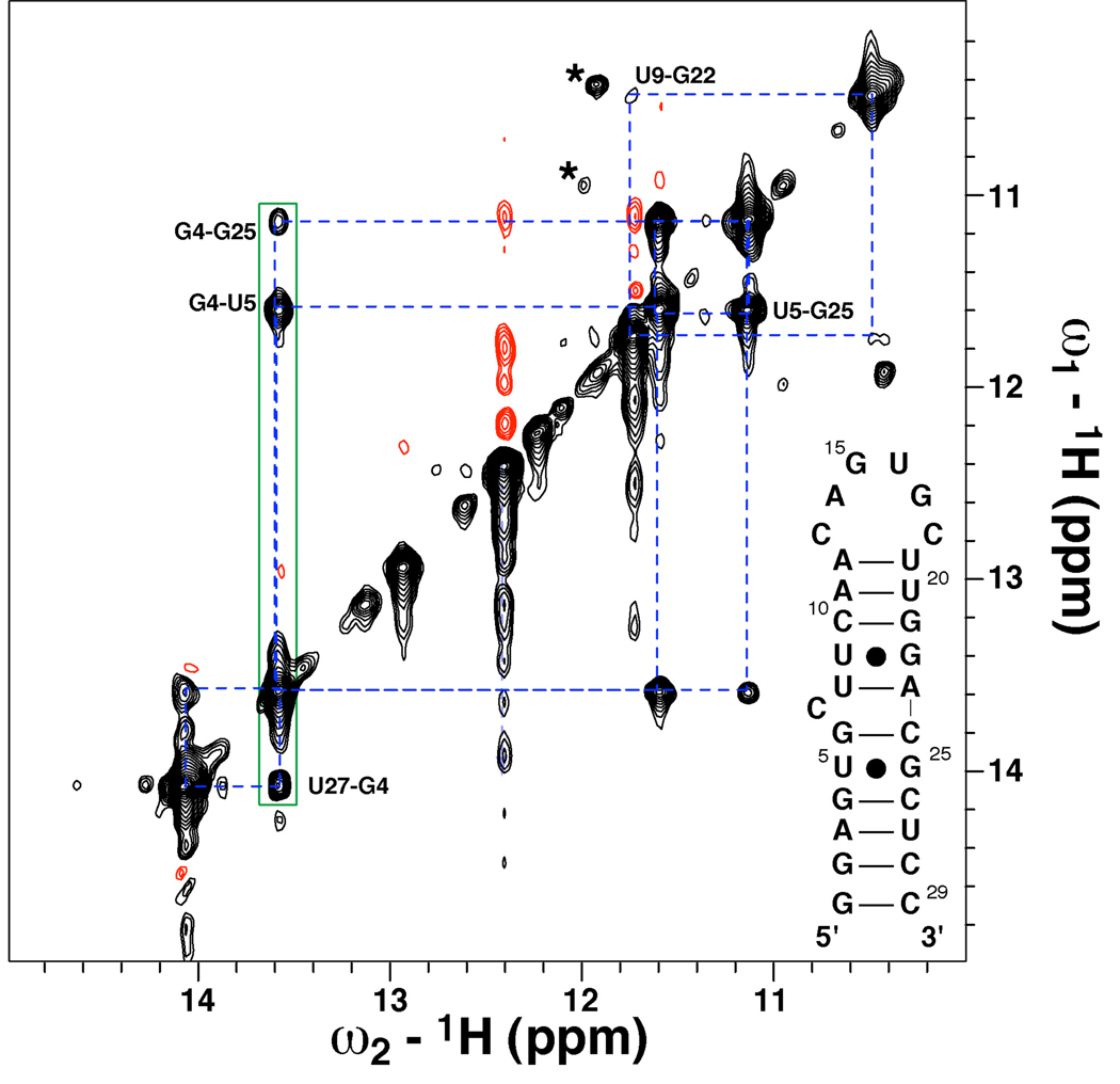
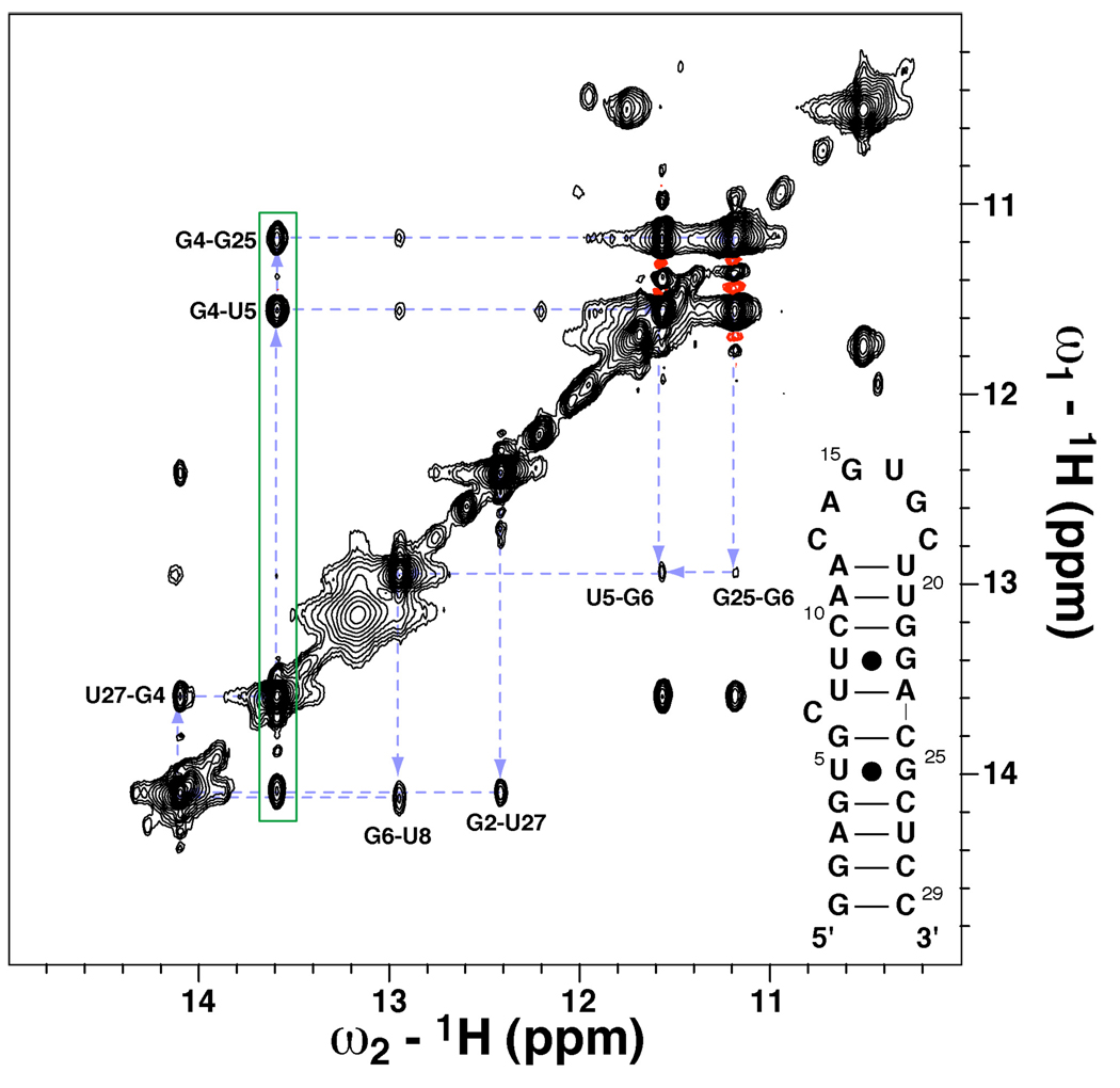
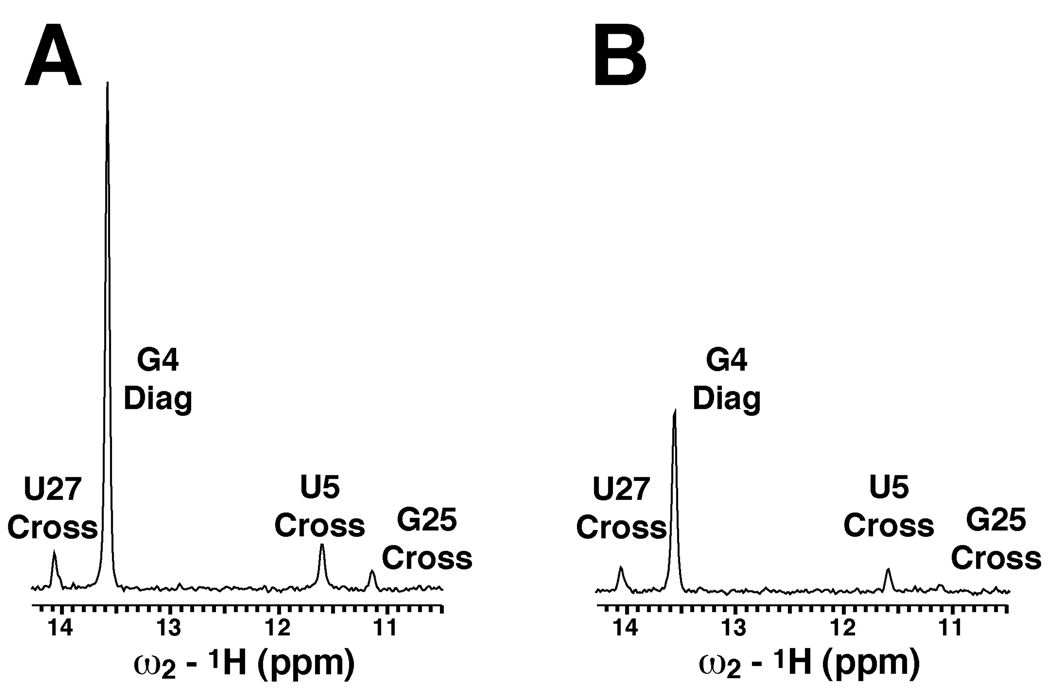
Imino (1)H-(15)N residual dipolar couplings (RDCs) provide additional structural information that complements standard (1)H-(1)H NOEs leading to improvements in both the local and global structure of RNAs. Here, we report measurement of imino (1)H-(1)H RDCs for the Iron Responsive Element (IRE) RNA and native E. coli tRNA(Val) using a BEST-Jcomp-HMQC2 experiment. (1)H-(1)H RDCs are observed between the imino protons in G-U wobble base pairs and between imino protons on neighboring base pairs in both RNAs. These imino (1)H-(1)H RDCs complement standard (1)H-(15)N RDCs because the (1)H-(1)H vectors generally point along the helical axis, roughly perpendicular to (1)H-(15)N RDCs. The use of longitudinal relaxation enhancement increased the signal-to-noise of the spectra by ~3.5-fold over the standard experiment. The ability to measure imino (1)H-(1)H RDCs offers a new restraint, which can be used in NMR domain orientation and structural studies of RNAs.


Figures

Figure 1
Figure 1
Imino region of the 2D 1H-1H BEST-Jcomp-HMQC2 experiment (Schanda, et al., 2007) shown in Supplemental Figure 1 collected on a 1 mM 15N-labeled IRE RNA containing ~17 mg/mL Pf1 bacteriophage. This spectrum was collected on a Varian INOVA 600 MHz spectrometer equipped with a cryogenically cooled triple resonance z-axis gradient probe operating at 25 °C. HMQC constant time delays were Δ1 = Δ2 = 35 ms. 140 × 2048 complex points were taken using sweep widths of 14006 Hz in the t1 and t2 dimensions. 400 scans per FID were collected with a 0.69 s inter-scan delay for a total experiment time of ~45 hrs. Black and red contours represent positive and negative intensity, respectively. The dashed blue lines illustrate the cross peaks which arise because of the transfer of magnetization due to imino 1H-1H RDCs. The asterisks denote cross peaks for the two G-U wobble base pairs, which are in an alternate conformation. The previously published imino assignments are indicated (Addess, et al., 1997), and the sequence and secondary structure of the IRE are shown in the lower right.
Figure 2
Figure 2
Plot of the absolute value of the experimental and predicted imino 1H-1H RDCs of the IRE. The predicted RDCs were obtained from the previously RDC-refined solution structure of the IRE. The experimental imino |DHH| values for data collected at 600 MHz (filled squares) are shown along with the average |DHH| values determined from triplicate measurements at 500 MHz (open circles). The predicted RDCs were obtained using an alignment tensor of DaNH = −18.8 Hz, R = 0.16, α = 24.1°, β = 108.4°, and γ = 123.7° and the refined solution structure (McCallum and Pardi, 2003) as input in REDCAT (Valafar and Prestegard, 2004). Assignments for the |DHH| interactions are given for each point, and error bars on the average |DHH| values represent the standard deviation calculated from triplicate experiments. The Pearson’s correlation coefficient, RP, average rmsd and Q-factor (Cornilescu, et al., 1998) for the 600 MHz data set are listed in the upper left-hand corner. These parameters were calculated without the |DHH| value for G22-U9 because of overlap of U9 and G21 imino proton resonances (see text).
Figure 3
Figure 3
Imino region of the 2D 1H-1H NOESY experiment with 175 ms mixing time collected on a 1 mM 15N-labeled IRE containing ~17 mg/mL Pf1 phage. This spectrum was collected at 5 °C on a Varian INOVA 600 MHz spectrometer equipped with a cryogenically cooled triple resonance z-axis probe. Water suppression was achieved with the gradient 11-echo scheme (Sklenár and Bax, 1987). Black and red contour lines represent positive and negative intensity, respectively. Dashed blue lines show the imino proton walk in the lower helix, and the green box highlights the three cross peaks to the imino proton of G4.
Figure 4
Figure 4
Experimentally observed imino DHH interactions are highlighted on the RDC-refined solution structure of the IRE (pdb entry 1NBR) (McCallum and Pardi, 2003). The alignment tensor (red axis system) resulting from alignment in ~17 mg/mL Pf1 bacteriophage is shown next to the structure (McCallum and Pardi, 2003). The lower stem containing some of the DHH interactions is expanded highlighting dipolar interactions for G4 labeled with their respective inter-nuclear distances.
Figure 5
Figure 5
1D traces for the G4 imino resonance taken from the 2D (A) BEST-Jcomp-HMQC2 and (B) SS-HMQC2 spectra on the IRE in Pf1 bacteriophage. Both spectra were collected at 500 MHz and 25 °C for ~20 hr, as described in the text. The assignments for G4 diagonal peak and U27, U5 and G25 cross peaks are given.

References

    1. Addess KJ, Basilion JP, Klausner RD, Rouault TA, Pardi A. Structure and dynamics of the iron responsive element RNA: Implications for binding of the RNA by iron regulatory binding proteins. J Mol Biol. 1997;274:72–83. - PubMed
    1. Al-Hashimi HM, Gosser Y, Gorin A, Hu WD, Majumdar A, Patel DJ. Concerted motions in HIV-1 TAR RNA may allow access to bound state conformations: RNA dynamics from NMR residual dipolar couplings. J Mol Biol. 2002;315:95–102. - PubMed
    1. Bax A, Kontaxis G, Tjandra N. Dipolar couplings in macromolecular structure determination. Methods Enzymol. 2001;339:127–174. - PubMed
    1. Boisbouvier J, Delaglio F, Bax A. Direct observation of dipolar couplings between distant protons in weekly aligned nucleic acids. Proc Natl Acad Sci USA. 2003;100:11333–11338. - PMC - PubMed
    1. Bondensgaard K, Mollova ET, Pardi A. The global conformation of the hammerhead ribozyme determined using residual dipolar couplings. Biochemistry. 2002;41:11532–11542. - PubMed

Publication types

MeSH terms