. 2008 Nov 25;6(11):e294.
doi: 10.1371/journal.pbio.0060294.

Prion switching in response to environmental stress

Affiliations

Prion switching in response to environmental stress

Jens Tyedmers et al. PLoS Biol. .

Abstract

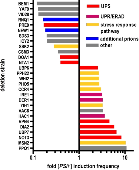
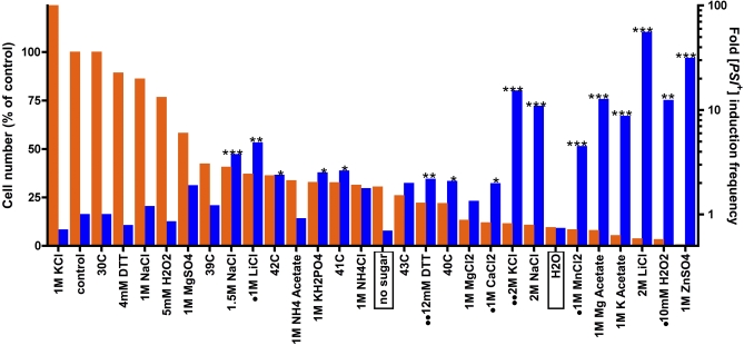
Evolution depends on the manner in which genetic variation is translated into new phenotypes. There has been much debate about whether organisms might have specific mechanisms for "evolvability," which would generate heritable phenotypic variation with adaptive value and could act to enhance the rate of evolution. Capacitor systems, which allow the accumulation of cryptic genetic variation and release it under stressful conditions, might provide such a mechanism. In yeast, the prion [PSI(+)] exposes a large array of previously hidden genetic variation, and the phenotypes it thereby produces are advantageous roughly 25% of the time. The notion that [PSI(+)] is a mechanism for evolvability would be strengthened if the frequency of its appearance increased with stress. That is, a system that mediates even the haphazard appearance of new phenotypes, which have a reasonable chance of adaptive value would be beneficial if it were deployed at times when the organism is not well adapted to its environment. In an unbiased, high-throughput, genome-wide screen for factors that modify the frequency of [PSI(+)] induction, signal transducers and stress response genes were particularly prominent. Furthermore, prion induction increased by as much as 60-fold when cells were exposed to various stressful conditions, such as oxidative stress (H2O2) or high salt concentrations. The severity of stress and the frequency of [PSI(+)] induction were highly correlated. These findings support the hypothesis that [PSI(+)] is a mechanism to increase survival in fluctuating environments and might function as a capacitor to promote evolvability.


Conflict of interest statement

Competing interests. The authors have declared that no competing interests exist.

Figures

Figure 1
Figure 1. A Genome-Wide Screen for Modifiers of [PSI +] Induction
(A) The intermediate toxicity caused by overexpressing the prion domain in [psi ] [RNQ +] provides an opportunity to screen for modifiers of [PSI +] induction. The prion domain (amino acids 1–253) of SUP35 was fused to YFP with an 8–amino acids (GSRVPVEK) linker (PD-YFP). Three different 74D-694 [RNQ +] derivatives express PD-YFP from genomic copies under control of the galactose-inducible promoter. The induction of PD-YFP on galactose medium is toxic in [PSI +] cells. In [psi ] cells partial toxicity is observed because only a fraction converts to [PSI +]. Confirming that [PSI +] induction is the cause of toxicity, cells carrying a SUP35 gene with its prion domain deleted (ΔPD) are immune. Serial dilutions were plated on SD (glucose) or SGal (galactose) medium and incubated for 3 d at 30°C. (B) Partial toxicity allowed us to identify both enhancers and suppressors of [PSI +] induction. We transformed 4,700 strains of the yeast gene deletion set (YGDS, Euroscarf) with a galactose-inducible PD-YFP 2μ plasmid in a high-throughput manner [67]. After selection for the plasmid, cells were spotted onto synthetic galactose-based medium to induce expression of PD-YFP. Some gene deletions cause a clear reduction of toxicity (decreased [PSI +] induction, exemplified here by hsp104Δ), whereas others further enhance toxicity (increased [PSI +] induction, exemplified by hac1Δ).
Figure 2
Figure 2. Large-Scale Secondary Screens to Retest Candidates
(A) Schematic overview and summary of large-scale testing approach to verify screen hits. (B) Construct specificity in candidates with increased toxicity. Candidate deletion strains that showed increased toxicity in the screen were re-arrayed in duplicate columns in 96-well plates. One column was transformed with the construct coding for galactose-inducible PD-YFP and the second column was transformed with a plasmid coding for galactose-inducible YFP. To induce overexpression of YFP or PD-YFP, cells were pinned onto galactose (inducing) medium and onto glucose (non-inducing) medium as a control. Effects in deletion strains were then compared to the parental BY4741 strain (BYwt) of the deletion library (YGDS). (C) Strategy to retest suppressors of toxicity. We used the strategy of SGA [45] to combine two copies of the galactose-inducible PD-YFP with our candidate deletions that suppressed toxicity. The donor strain ([RNQ +]) was mated to the candidate gene deletion strains by pinning onto plates containing the appropriate complete synthetic medium (SD). After overnight incubation, we replica-plated them four times onto medium selective for diploids. Diploids were then sporulated in liquid medium in 96-well plates at 23 °C for 6 d. Sporulation cultures were pinned and replica-plated three times onto selection plates for spores that combine the desired features. Overexpression of PD-YFP was achieved by spotting onto galactose medium, or glucose-based medium as a control.
Figure 3
Figure 3. Measuring Spontaneous [PSI +] Induction Frequency
Candidate genes were knocked out in the repeat expansion strain 74D-694-R2E2 [28]. Cultures of different knockout strains and wild-type strains (150 μl for each) without any knockout were grown in 96-well plates for 48 h at 30 °C on a shaker. Each culture (12.5 μl) was plated directly onto adenine-deficient SD plates (SD-Ade), and a 1:500 dilution of the culture was plated onto complete SD plates as a control. Complete SD plates were incubated for 3 d at 30 °C and SD-Ade plates were incubated for 7 d at 30 °C. Colonies on the plates were counted using an Acolyte colony counter. Colonies on SD-Ade plates were tested for curability [50] by replica plating onto plates containing 3 mM guanidine HCl and subsequent reprobing on SD-Ade plates. Non-curable colonies were subtracted. For gene deletion strains, all ADE+ colonies were tested for curability, whereas for stress conditions, a representative subset was tested. Curability was always above 95%. Relative induction frequencies were calculated by dividing the number of colonies on SD-Ade plates by the number on complete synthetic medium.
Figure 4
Figure 4. Gene Deletions That Affect [PSI +] Induction Frequency
Strains that were 74D-694-R2E2 [psi ] [RNQ +] [28] and that carried the 40 deletions that were tested most thoroughly were cured of any [PSI +] elements that might have appeared spontaneously by growing cells carrying a 2μ plasmid carrying HSP104 regulated by a galactose-inducible promoter [18]. Cells were transferred to glucose medium in 96-well plates and grown for 48 h at 30°C. [PSI +] induction frequency was determined first by plating aliquots onto selective medium (SD-Ade) followed by testing for curability by guanidine HCl, which is known to eliminate [PSI +] [50] (compare Figure 3). The data shown are the results of seven independent experiments testing a total of at least six independent transformants for each deletion strain. The least square mean values of [PSI +] induction frequency (p < 0.05 to p < 0.001) were plotted after normalizing their values to the wild type. The bars are coded to indicate different functional groups: UPS, ubiquitin-proteasome system; UPR, unfolded protein response; ERAD, endoplasmic-reticulum-associated degradation.
Figure 5
Figure 5. Stressful Growth Conditions Trigger Induction of the [PSI +] Prion
A culture of 74D-694-R2E2 was transferred to a 96-well plate at an optical density of 0.25 and incubated with complete synthetic medium as a control or medium supplemented with the indicated substances and incubated for 12–24 h at 30°C or higher temperatures as indicated. Cultures were subsequently plated on complete synthetic medium (SD) and on medium selective for [PSI +] (SD-Ade) to determine the frequency of [PSI +] induction (Figure 3). [PSI +] status was confirmed by guanidine HCl curing. Orange bars, cell numbers determined on complete synthetic medium (left axis). Blue bars, [PSI +] induction frequency (right axis). Conditions that significantly increased [PSI +] induction relative to control are indicated by asterisks (*p < 0.05, **p < 0.01, ***p < 0.001). [PSI +] cells had a growth disadvantage in conditions indicated by dots and had a growth advantage in conditions indicated by double dots. Significant increases in [PSI +] induction frequency were determined as described in Materials and Methods.

References

    1. Cox B. [PSI], a cytoplasmic suppressor of super-suppression in yeast. Heredity. 1965;20:505–521.
    1. Liebman SW, Sherman F. Extrachromosomal psi+ determinant suppresses nonsense mutations in yeast. J Bacteriol. 1979;139:1068–1071. - PMC - PubMed
    1. Tuite MF, Cox BS. Propagation of yeast prions. Nat Rev Mol Cell Biol. 2003;4:878–890. - PubMed
    1. Shorter J, Lindquist S. Prions as adaptive conduits of memory and inheritance. Nat Rev Genet. 2005;6:435–450. - PubMed
    1. Chien P, Weissman JS, DePace AH. Emerging principles of conformation-based prion inheritance. Annu Rev Biochem. 2004;73:617–656. - PubMed

Publication types

MeSH terms