Review
. 2009 Feb;100(2):209-15.
doi: 10.1111/j.1349-7006.2008.01034.x.

MEN1 gene and its mutations: basic and clinical implications

Affiliations
Review

MEN1 gene and its mutations: basic and clinical implications

Toshihiko Tsukada et al. Cancer Sci. 2009 Feb.

Abstract

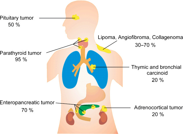
Heterozygous germline mutations of the tumor-suppressor gene MEN1 are responsible for multiple endocrine neoplasia type 1 (MEN1), a dominantly inherited familial cancer syndrome characterized by pituitary, parathyroid, and enteropancreatic tumors. Various mutations have been identified throughout the entire gene region in patients with MEN1 and related disorders. Neither mutation hot spot nor phenotype–genotype correlation has been established in MEN1 although some missense mutations may be specifically associated with a phenotype of familial isolated hyperparathyroidism. The gene product menin has been implicated in multiple roles, including gene transcription, maintenance of genomic integrity, and control of cell division and differentiation. These multiple functions are likely to be conferred by association with multiple protein factors. Occurrence of MEN1-causing missense mutations throughout menin also suggests the requirement of multiple binding factors for its full tumor-suppressive activity. The effect of menin depletion is highly tissue specific, but its underlying mechanism remains to be elucidated. A DNA test for MEN1 germline mutations is a useful tool for diagnosis of MEN1 although it needs further improvements


Figures

Figure 1
Figure 1
Estimated penetrance of tumors arising in MEN1.
Figure 2
Figure 2
Chromosomal localization and structural organization of the human MEN1 gene. (a) Chromosome 11. (b) The approximately 300‐kb genomic region containing the MEN1 gene (closed box) and microsatellites (red bar) used as polymorphic DNA markers. Cen, centromere; Tel, telomere. (c) MEN1 gene. Green and yellow boxes indicate protein‐coding and non‐coding regions of exons, respectively. (d) Menin mRNA. Green and yellow boxes indicate translated and untranslated regions of the mRNA, respectively. (e) Menin. Blue regions indicate nuclear localization signals.
Figure 3
Figure 3
Mutations, evolutionary diversion, and binding domains of mein. (a) Germline MEN1 mutations identified in multiple endocrine neoplasia type 1 (MEN1) and related disorders. Locations of missense mutations (perpendicular lines), in‐frame deletions (dot, one amino acid; horizontal bar, two or more amino acids), and in‐frame insertions (triangles) are shown above the diagram of menin, with corresponding exons numbered. Green symbols indicate mutations causing MEN1, and orange symbols indicate those causing familial isolated hyperparathyroidism or apparently sporadic parathyroid tumor. Frameshift mutations (open triangles) and nonsense mutations (closed triangles) are shown below menin, with potential nonsense mutation sites indicated by dots. Splicing mutations and large deletions are not depicted. Blue boxes indicate nuclear localization signals. The normal polymorphisms R171Q and A541T are also indicated. #Nonsense mutation involving two base pair substitutions. (b) Homology between menin amino acid sequences among different vertebrate species. Sequences were retrieved from the internet website: http://www.ncbi.nlm.nih.gov/HomoloGene/. Blue and red dots indicate conservative and non‐conservative amino acid replacement compared with human menin, respectively. Red triangles indicate insertions of amino acid residues. (c) Menin regions implicated in its binding to interacting proteins. Blue bars represent the regions required for binding to proteins demonstrated to bind directly to menin. Green bars indicate the regions required for association with proteins not shown to bind directly to menin.
Figure 4
Figure 4
Postulated roles for menin in transcriptional repression and activation. In this model, menin represses pro‐cell growth genes by recruiting a general corepressor mSin3A and histone deacetylase (HDAC) to specific transcription factor (TF) binding sites, thus promoting histone deacetylation (upper). On the other hand, menin activates transcription of genes involved in cell growth inhibition and cell differentiation by recruiting lens epithelium‐derived growth factor (LEDGF) and histone methyltransferase (HMT) to specific TF binding sites, thus promoting histone methylation (lower).

References

    1. Chandrasekharappa SC, Guru SC, Manickam P et al . Positional cloning of the gene for multiple endocrine neoplasia‐type 1. Science 1997; 276: 404–7. - PubMed
    1. Gardner DG. Recent advances in multiple endocrine neoplasia syndromes. Adv Intern Med 1997; 42: 597–627. - PubMed
    1. Brandi ML, Gagel RF, Angeli A et al . Guidelines for diagnosis and therapy of MEN type 1 and type 2. J Clin Endocrinol Metab 2001; 86: 5658–71. - PubMed
    1. Lemos MC, Thakker RV. Multiple endocrine neoplasia type 1 (MEN1): analysis of 1336 mutations reported in the first decade following identification of the gene. Hum Mutat 2008; 29: 22–32. - PubMed
    1. Balogh K, Hunyady L, Patocs A et al . MEN1 gene mutations in Hungarian patients with multiple endocrine neoplasia type 1. Clin Endocrinol 2007; 67: 727–34. - PubMed

Publication types

MeSH terms

Substances