. 2008 Dec 10:8:216.
doi: 10.1186/1471-2180-8-216.

Acinetobacter baumannii invades epithelial cells and outer membrane protein A mediates interactions with epithelial cells

Affiliations

Acinetobacter baumannii invades epithelial cells and outer membrane protein A mediates interactions with epithelial cells

Chul Hee Choi et al. BMC Microbiol. .

Abstract

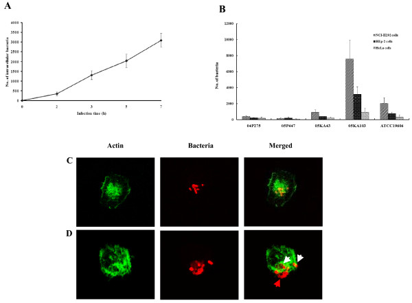
Background: Acinetobacter baumannii is a nosocomial pathogen of increasing importance, but the pathogenic mechanism of this microorganism has not been fully explored. This study investigated the potential of A. baumannii to invade epithelial cells and determined the role of A. baumannii outer membrane protein A (AbOmpA) in interactions with epithelial cells.

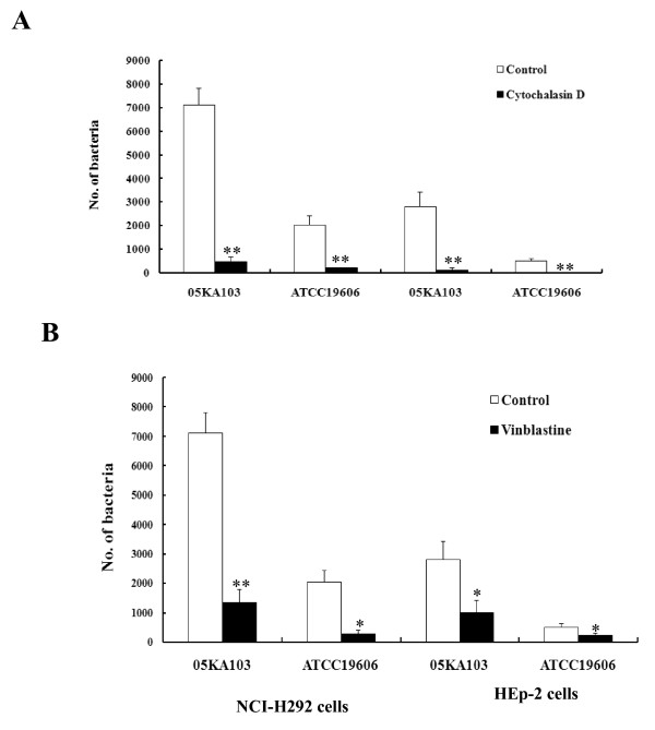
Results: A. baumannii invaded epithelial cells by a zipper-like mechanism, which is associated with microfilament- and microtubule-dependent uptake mechanisms. Internalized bacteria were located in the membrane-bound vacuoles. Pretreatment of recombinant AbOmpA significantly inhibited the adherence to and invasion of A. baumannii in epithelial cells. Cell invasion of isogenic AbOmpA- mutant significantly decreased as compared with wild-type bacteria. In a murine pneumonia model, wild-type bacteria exhibited a severe lung pathology and induced a high bacterial burden in blood, whereas AbOmpA- mutant was rarely detected in blood.

Conclusion: A. baumannii adheres to and invades epithelial cells. AbOmpA plays a major role in the interactions with epithelial cells. These findings contribute to the understanding of A. baumannii pathogenesis in the early stage of bacterial infection.


Figures

Figure 1
Figure 1
Invasion of A. baumannii in epithelial cells. (A) NCI-H292 cells were infected with A. baumannii ATCC 19606T at an MOI of 100 up to 7 h. The colony-forming units were enumerated to measure the time-course of invasion. The result represents the mean ± standard deviation in duplicate wells and repeated a minimum of three separate times on separate days. (B) NCI-H292, HEp-2, and HeLa cells were infected with A. baumannii strains at an MOI of 100 for 5 h. (C) NCI-H292 cells were infected with A. baumannii 05KA103 at an MOI of 100 for 5 h. Actin was stained with Alexa Fluor ® 488 phalloidin (green). Bacteria were stained with polyclonal anti-rabbit AbOmpA antibody, followed by a secondary antibody Alexa Fluor ® 568 (red). The analytical sectioning was performed from the top to the bottom of the cells. The figure represents a single section of the cells. (D). NCI-H292 cells were infected with A. baumannii ATCC 19606T at an MOI of 100 for 5 h. Actin filaments have wrap-around-bacteria (white arrow). Red arrow indicates the extracellular bacteria. The figure represents all projection of sections in one picture.
Figure 2
Figure 2
Inhibition of A. baumannii invasion by cytoskeleton inhibitors, cytochalasin D (A) and vinblastine (B). NCI-H292 and HEp-2 cells were pretreated with cytochalasin D (2 μM) or vinblastine (100 μM) for 30 min and maintained for 5 h of the entire infection period. A. baumannii invasion was measured as the number of bacteria per well using the gentamicin protection assay. Data represent the mean ± standard deviation of three separate experiments. *P < 0.01, **P < 0.005.
Figure 3
Figure 3
Electron micrographs demonstrating internalization of A. baumannii in epithelial cells. NCI-H292 cells were infected with A. baumannii 05KA103 at an MOI of 100 for 5 h. (A and B) SEM images showing interaction of A. baumannii with NCI-H292 cells. The cell membrane was extended to and wrapped around A. baumannii (arrow). Bar represents 2 μm. (C to E) TEM images. (C) A. baumannii loosely anchored to the cell surface (arrow) and small protrusion from the cell was appeared (arrow head). (D) The cell membrane was fused after the bacterial internalization (arrow). (E) Intracellular bacteria were surrounded by a membrane-bound vacuole (arrow head). Bar represents 1 μm.
Figure 4
Figure 4
Cell surface binding of rAbOmpA. The grey shaded region represents the control fluorescent level seen in the cells treated with polyclonal anti-rabbit AbOmpA antibody and Alexa Fluor ® 488-conjugated secondary antibody without the addition of rAbOmpA. The solid line represents the fluorescent level seen in the cells treated with 6 μg/ml of rAbOmpA.
Figure 5
Figure 5
rAbOmpA attenuates the interaction of A. baumannii with epithelial cells. (A) NCI-H292 cells were pretreated with 10 μg/ml of rAbOmpA for 1 h and then infected with A. baumannii 05KA103 at an MOI of 100 for 1 h. Cell monolayer was stained with the Giemsa solution. (B) NCI-H292 and HEp-2 cells were pretreated with 10 μg/ml of rAbOmpA for 1 h and infected with A. baumannii at an MOI of 100 for 5 h. Invasion efficiency was expressed as the number of bacteria per well using the gentamicin protection assay. The results represent the mean ± standard deviation of three separate experiments. *P < 0.05.
Figure 6
Figure 6
Adherence and invasion of A. baumannii ATCC19606T and isogenic AbOmpA- mutant in epithelial cells. (A). Adherence of A. baumannii to epithelial cells. NCI-H292 cells were infected with A. baumannii strains at an MOI of 100 for 1 h. Actin was stained with Alexa Fluor ® 488 phalloidin (green). Bacteria were stained with polyclonal anti-rabbit AbOmpA antibody, followed by a secondary antibody Alexa Fluor ® 568 (red). (B). NCI-H292 and HEp-2 cells were infected with A. baumannii at an MOI of 100 for 5 h. Invasion efficiency was expressed as the number of bacteria per well using the gentamicin protection assay. Results represent the mean and standard deviation of three separate experiments on separately days. *P < 0.001.

References

    1. Dijkshoorn L, Nemec A, Seifert H. An increasing threat in hospitals: multidrug-resistant Acinetobacter baumannii. Nat Rev Microbiol. 2007;5:939–951. doi: 10.1038/nrmicro1789. - DOI - PubMed
    1. Peleg AY, Seifert H, Paterson DL. Acinetobacter baumannii: emergence of a successful pathogen. Clin Microbiol Rev. 2008;21:538–582. doi: 10.1128/CMR.00058-07. - DOI - PMC - PubMed
    1. Go ES, Urban C, Burns J, Kreiswirth B, Eisner W, Mariano N, Mosinka-Snipas K, Rahal JJ. Clinical and molecular epidemiology of Acinetobacter infections sensitive only to polymyxin B and sulbactam. Lancet. 1994;344:1329–1332. doi: 10.1016/S0140-6736(94)90694-7. - DOI - PubMed
    1. Seifert H, Strate A, Schulze A, Pulverer G. Bacteremia due to Acinetobacter species other than Acinetobacter baumannii. Infection. 1994;22:379–385. doi: 10.1007/BF01715492. - DOI - PubMed
    1. Nemec A, Dijkshoorn L, Cleenwerck I, De Baere T, Janssens D, Reijden TJ van der, Jezek P, Vaneechoutte M. Acinetobacter parvus sp. Nov., a small-colony-forming species isolated from human clinical specimens. Int J Sys Evol Microbiol. 2003;53:1563–1567. doi: 10.1099/ijs.0.02631-0. - DOI - PubMed

Publication types

MeSH terms

LinkOut - more resources