. 2008 Nov;83(5):643-8.
doi: 10.1016/j.ajhg.2008.10.010.

Mutations in the fatty acid 2-hydroxylase gene are associated with leukodystrophy with spastic paraparesis and dystonia

Affiliations

Mutations in the fatty acid 2-hydroxylase gene are associated with leukodystrophy with spastic paraparesis and dystonia

Simon Edvardson et al. Am J Hum Genet. 2008 Nov.

Abstract

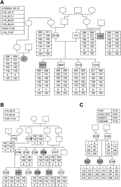
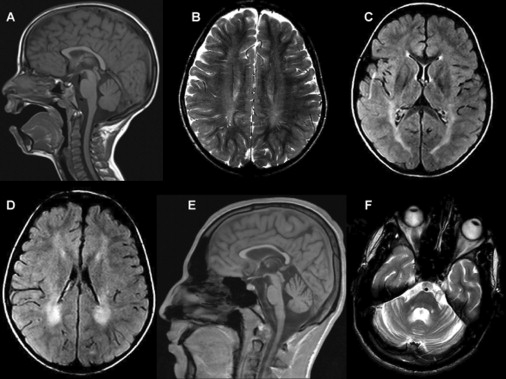
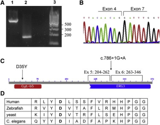
Myelination is a complex, developmentally regulated process whereby myelin proteins and lipids are coordinately expressed by myelinating glial cells. Homozygosity mapping in nine patients with childhood onset spasticity, dystonia, cognitive dysfunction, and periventricular white matter disease revealed inactivating mutations in the FA2H gene. FA2H encodes the enzyme fatty acid 2-hydroxylase that catalyzes the 2-hydroxylation of myelin galactolipids, galactosylceramide, and its sulfated form, sulfatide. To our knowledge, this is the first identified deficiency of a lipid component of myelin and the clinical phenotype underscores the importance of the 2-hydroxylation of galactolipids for myelin maturation. In patients with autosomal-recessive unclassified leukodystrophy or complex spastic paraparesis, sequence analysis of the FA2H gene is warranted.


Figures

Figure 1
Figure 1
The Families' Pedigree and Haplotype along the Critical Region on Chromosome 16 (A) Family 1 pedigree and their haplotype along the critical region on chromosome 16. Patients' symbols are filled. Numbered symbols represent individuals whose DNA samples were available for analysis. The polymorphic microsatellite markers and their chromosomal locations (in Mb) are given in the upper-left panel. C16 _65.77 stands for hg18_chr16:65767359-65767624, C16_67.21 stands for hg18_chr16:67214180-67214437, C16_68.20 stands for hg18_ chr16:68218628-68218887, C16_69.20 stands for hg18_chr16:69227890-69228133, and C16_73.87 stands for hg18_chr16:73875745-73875997. (B) Family 2 pedigree. The asterisk represents an individual whose family name is identical to that of family 1. (C) Family 3 pedigree. The polymorphic microsatellite markers and their location are given at the upper-left and upper-right panel, for family 2 and family 3, respectively.
Figure 2
Figure 2
Electron Micrograph of the Sural Nerve Showing Normal Structure and Distribution of Myelinated and Unmyelinated Nerve Fibers The inset shows details of a myelin sheath to show normal compaction and periodicity of myelin lamellae.
Figure 3
Figure 3
Brain MRI of Patient 2762 at 7 Years of Age and Patient 1211 at 18 Years of Age (A) Sagittal T1 weighted image. (B) Axial T2 weighted image. (C and D) Axial FLAIR images showing prolonged relaxation times in the periventricular white matter, more prominent posteriorly, and to a lesser extent in the posterior limbs of the internal capsules. There is no significant loss of volume. Images in (A)–(D) are of patient 2762 at 7 years of age. (E and F) Sagittal T1 weighted (E) and axial T2 weighted (F) images showing loss of volume of the corpus callosum and to a lesser extent of the cerebellum and pons. Images are of patient 1211 at 18 years of age.
Figure 4
Figure 4
Mutations in the FA2H Gene (A) A cDNA fragment of the FA2H gene from the patients of family 1 and 2 who are homozygous for the c.786+1G→A mutation. Lane 1 represents the control, lane 2 represents the patient, and lane 3 represents the DNA ladder marker. (B) Sequence of the short cDNA fragment demonstrating exon 5 and 6 skipping. (C) Schematic representation of the conserved domains of the FA2H protein. Cyt-b5 stands for cytochrome b5-like heme-binding domain and ERG3 is the sterol desaturase domain. The locations of the residues encoded by exon 5 and 6 and of the two mutations are shown. (D) Conservation throughout evolution of the aspartic acid at codon 35 (in bold), which is mutated in patients from family 3.

References

    1. Morell P., Quarles R.H. Myelin Formation, Structure and Biochemistry. In: Siegel G.J., Agranoff B.W., Fisher S.K., Albers R.W., Uhler M.D., editors. Basic Neurochemistry. Molecular, Cellular and Medical Aspects. Sixth Edition. Lippincott-Raven; New York: 1999. pp. 69–94.
    1. Campagnoni A.T. Molecular biology of myelination. In: Kettenmann H., Ransom B.R., editors. Neuroglia. Oxford University Press; New York: 2004. pp. 253–263.
    1. Norton W.T., Cammer W. Isolation and characterization of myelin. In: Morell P., editor. Myelin. Plenum Press; New York: 1984. pp. 147–195.
    1. Webster H.D.F., Sternberger N.H. Morphological features of myelin formation. In: Bauman N., editor. Neurological mutations affecting myelination. INSERM Symposium. Vol. 14. Elsevier; 1980. pp. 73–86.
    1. Edvardson S., Shaag S., Kolesnikova O., Gomori J.M., Tarassov I., Einbinder T., Saada A., Elpeleg O. Deleterious mutation in the mitochondrial arginyl-tRNA synthetase gene is associated with ponto-cerebellar hypoplasia. Am. J. Hum. Genet. 2007;81:857–862. - PMC - PubMed

Publication types

MeSH terms

Substances