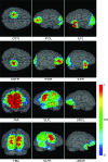
Mapping of functional areas in the human cortex based on connectivity through association fibers
- PMID: 19068488
- PMCID: PMC2705697
- DOI: 10.1093/cercor/bhn215
Mapping of functional areas in the human cortex based on connectivity through association fibers
Abstract
In the human brain, different regions of the cortex communicate via white matter tracts. Investigation of this connectivity is essential for understanding brain function. It has been shown that trajectories of white matter fiber bundles can be estimated based on orientational information that is obtained from diffusion tensor imaging (DTI). By extrapolating this information, cortical regions associated with a specific white matter tract can be estimated. In this study, we created population-averaged cortical maps of brain connectivity for 4 major association fiber tracts, the corticospinal tract (CST), and commissural fibers. It is shown that these 4 association fibers interconnect all 4 lobes of the hemispheres. Cortical regions that were assigned based on association with the CST and the superior longitudinal fasciculus (SLF) agreed with locations of their known (CST: motor) or putative (SLF: language) functions. The proposed approach can potentially be used for quantitative assessment of the effect of white matter abnormalities on associated cortical regions.
Figures


Similar articles
-
White matter tractography using diffusion tensor deflection.Hum Brain Mapp. 2003 Apr;18(4):306-21. doi: 10.1002/hbm.10102. Hum Brain Mapp. 2003. PMID: 12632468 Free PMC article.
-
Characterization of displaced white matter by brain tumors using combined DTI and fMRI.Neuroimage. 2006 May 1;30(4):1100-11. doi: 10.1016/j.neuroimage.2005.11.015. Epub 2006 Jan 19. Neuroimage. 2006. PMID: 16427322
-
Development of cerebral fiber pathways in cats revealed by diffusion spectrum imaging.Neuroimage. 2010 Jan 15;49(2):1231-40. doi: 10.1016/j.neuroimage.2009.09.002. Epub 2009 Sep 8. Neuroimage. 2010. PMID: 19747553 Free PMC article.
-
Diffusion tensor tractography of the motor white matter tracts in man: Current controversies and future directions.Ann N Y Acad Sci. 2005 Dec;1064:88-97. doi: 10.1196/annals.1340.016. Ann N Y Acad Sci. 2005. PMID: 16394150 Review.
-
[White matter fiber tractography and quantitative analysis of diffusion tensor imaging].Brain Nerve. 2015 Apr;67(4):475-85. doi: 10.11477/mf.1416200164. Brain Nerve. 2015. PMID: 25846596 Review. Japanese.
Cited by
-
Causality in the association between P300 and alpha event-related desynchronization.PLoS One. 2012;7(4):e34163. doi: 10.1371/journal.pone.0034163. Epub 2012 Apr 12. PLoS One. 2012. PMID: 22511933 Free PMC article.
-
White matter abnormalities in paediatric obsessive-compulsive disorder: a systematic review of diffusion tensor imaging studies.Brain Imaging Behav. 2023 Jun;17(3):343-366. doi: 10.1007/s11682-023-00761-x. Epub 2023 Mar 20. Brain Imaging Behav. 2023. PMID: 36935464 Free PMC article.
-
fMRI-DTI modeling via landmark distance atlases for prediction and detection of fiber tracts.Neuroimage. 2012 Mar;60(1):456-70. doi: 10.1016/j.neuroimage.2011.11.014. Epub 2011 Dec 2. Neuroimage. 2012. PMID: 22155376 Free PMC article.
-
Language in schizophrenia: relation with diagnosis, symptomatology and white matter tracts.NPJ Schizophr. 2020 Apr 20;6(1):10. doi: 10.1038/s41537-020-0099-3. NPJ Schizophr. 2020. PMID: 32313047 Free PMC article.
-
Three-way (N-way) fusion of brain imaging data based on mCCA+jICA and its application to discriminating schizophrenia.Neuroimage. 2013 Feb 1;66:119-32. doi: 10.1016/j.neuroimage.2012.10.051. Epub 2012 Oct 26. Neuroimage. 2013. PMID: 23108278 Free PMC article.
References
-
- Basser PJ, Mattiello J, LeBihan D. Estimation of the effective self-diffusion tensor from the NMR spin echo. J Magn Reson B. 1994a;103:247–254. - PubMed
-
- Basser PJ, Pajevic S, Pierpaoli C, Duda J, Aldroubi A. In vitro fiber tractography using DT-MRI data. Magn Reson Med. 2000;44:625–632. - PubMed
-
- Carpenter M. Human neuroanatomy. Baltimore (MD): Williams & Wilkins; 1976.
-
- Catani M, Howard RJ, Pajevic S, Jones DK. Virtual in vivo interactive dissection of white matter fasciculi in the human brain. Neuroimage. 2002;17:77–94. - PubMed

