

Review
doi: 10.1016/j.exer.2008.11.007.
Epub 2008 Nov 24.
Lens intermediate filaments
Affiliations
- PMID: 19071112
- PMCID: PMC2696462
- DOI: 10.1016/j.exer.2008.11.007
Item in Clipboard
Review
Lens intermediate filaments
Exp Eye Res.
2009 Feb.
Abstract
The ocular lens assembles two separate intermediate filament systems sequentially with differentiation. Canonical 8-11 nm IFs composed of Vimentin are assembled in lens epithelial cells and younger fiber cells, while the fiber cell-specific beaded filaments are switched on as fiber cell elongation initiates. Some of the key features of both filament systems are reviewed.
Figures
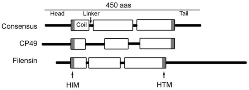
Schematic view of the consensus predicted secondary structure of the IF protein, along with that predicted for CP49 and filensin.
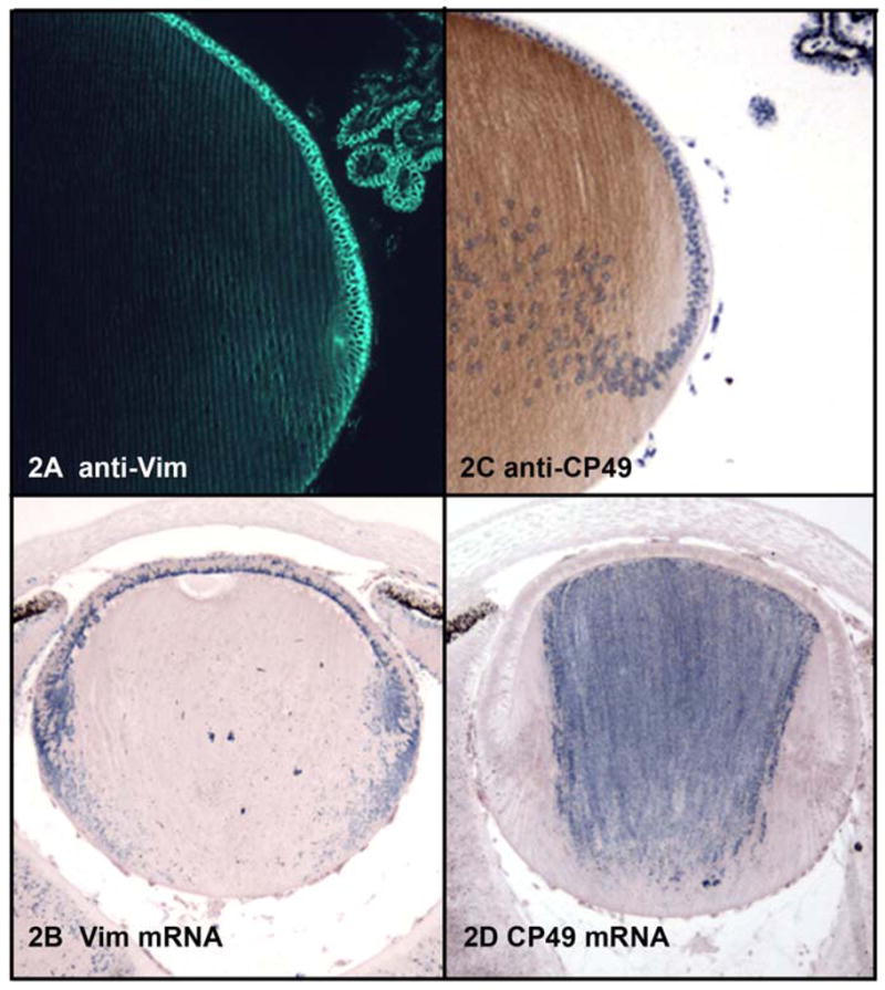
2A: Immunoflurescent localization of vimentin in a paraffin section of mouse lens fixed by freeze substitution in 97% methanol:3% acetic acid. 2B: In situ hybridization localizing vimentin mRNA in paraffin sections of mouse lens. 2C: Immunoperoxidase localization of CP49 in paraffin section of mouse lens, fixed as in 2A. 2D: In situ hybridization demonstrating distribution of CP49 mRNA in mouse lens.
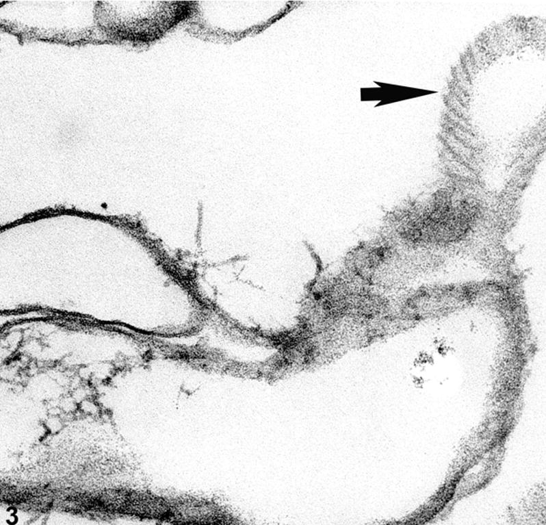
Electron micrograph of ghosted mouse lens, showing a tangential view of a fiber cell membrane (arrow) where IFs are aligned along the surface of the membrane.
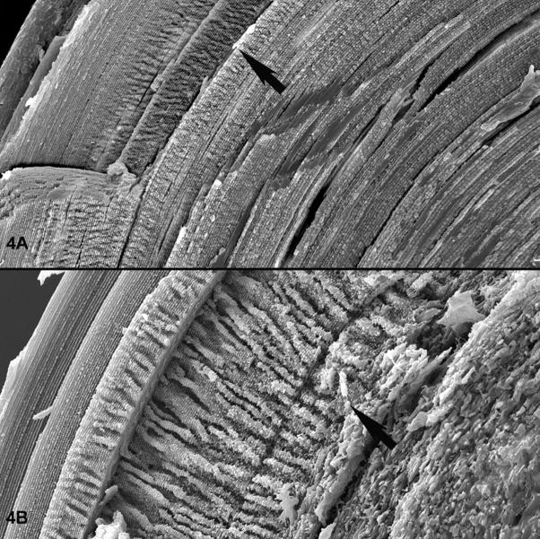
Scanning electron micrographs of a wild type (A) and BF knockout (B) lens. Figure A is a lower magnification overview, with an arrow highlighting the transition from a region where the paddle-like structures are regressing. Deep to this point, fiber cells retain their unique shape and strict alignment. Figure B is a higher magnification view of a BF knockout lens, with an arrow placed at a similar transition point. The loss of fiber cell shape and alignment at this juncture is evident in the knockout.
Similar articles
-
Development- and differentiation-dependent reorganization of intermediate filaments in fiber cells.Invest Ophthalmol Vis Sci. 2001 Mar;42(3):735-42. Invest Ophthalmol Vis Sci. 2001. PMID: 11222535
-
The beaded intermediate filaments and their potential functions in eye lens.Bioessays. 1994 Jun;16(6):413-8. doi: 10.1002/bies.950160609. Bioessays. 1994. PMID: 8080431 Review.
-
Filensin and phakinin form a novel type of beaded intermediate filaments and coassemble de novo in cultured cells.J Cell Biol. 1996 Feb;132(4):643-55. doi: 10.1083/jcb.132.4.643. J Cell Biol. 1996. PMID: 8647895 Free PMC article.
-
Periplakin interactions with lens intermediate and beaded filaments.Invest Ophthalmol Vis Sci. 2009 Mar;50(3):1283-9. doi: 10.1167/iovs.08-2894. Epub 2008 Nov 21. Invest Ophthalmol Vis Sci. 2009. PMID: 19029034 Free PMC article.
-
Seeing is believing! The optical properties of the eye lens are dependent upon a functional intermediate filament cytoskeleton.Exp Cell Res. 2005 Apr 15;305(1):1-9. doi: 10.1016/j.yexcr.2004.11.021. Epub 2005 Jan 26. Exp Cell Res. 2005. PMID: 15777782 Review.
Cited by
-
Role of Aquaporin 0 in lens biomechanics.Biochem Biophys Res Commun. 2015 Jul 10;462(4):339-45. doi: 10.1016/j.bbrc.2015.04.138. Epub 2015 May 8. Biochem Biophys Res Commun. 2015. PMID: 25960294 Free PMC article.
-
Localization of the lens intermediate filament switch by imaging mass spectrometry.Exp Eye Res. 2020 Sep;198:108134. doi: 10.1016/j.exer.2020.108134. Epub 2020 Jul 16. Exp Eye Res. 2020. PMID: 32682822 Free PMC article.
-
Ultrastructural analysis of the human lens fiber cell remodeling zone and the initiation of cellular compaction.Exp Eye Res. 2013 Nov;116:411-8. doi: 10.1016/j.exer.2013.10.015. Epub 2013 Oct 30. Exp Eye Res. 2013. PMID: 24183661 Free PMC article.
-
Lens Biology and Biochemistry.Prog Mol Biol Transl Sci. 2015;134:169-201. doi: 10.1016/bs.pmbts.2015.04.007. Epub 2015 Jun 4. Prog Mol Biol Transl Sci. 2015. PMID: 26310155 Free PMC article. Review.
-
CD24 is required for sustained transparency of the adult lens.Exp Eye Res. 2025 Jun;255:110347. doi: 10.1016/j.exer.2025.110347. Epub 2025 Mar 18. Exp Eye Res. 2025. PMID: 40112946
References
-
- Achstatter T, Moll R, et al. Expression of glial filament protein (GFP) in nerve sheaths and non-neural cells re-examined using monoclonal antibodies, with special emphasis on the co-expression of GFP and cytokeratins in epithelial cells of human salivary gland and pleomorphic adenomas. Differentiation. 1986;31(3):206–27. - PubMed
-
- Agbulut O, Li Z, et al. Analysis of skeletal and cardiac muscle from desmin knock-out and normal mice by high resolution separation of myosin heavy-chain isoforms. Biol Cell. 1996;88(3):131–5. - PubMed
-
- Albers K, Fuchs E. The molecular biology of intermediate filament proteins. Int Rev Cytol. 1992;134:243–79. - PubMed
-
- Alizadeh A, Clark J, et al. Targeted deletion of the lens fiber cell-specific intermediate filament protein filensin. Invest Ophthalmol Vis Sci. 2003;44(12):5252–8. - PubMed
-
- Alizadeh A, Clark J, et al. Characterization of a mutation in the lens-specific CP49 in the 129 strain of mouse. Invest Ophthalmol Vis Sci. 2004;45(3):884–91. - PubMed
Publication types
MeSH terms
Substances
Grants and funding
LinkOut - more resources
Full Text Sources
