. 2009 Apr;44(4):708-16.
doi: 10.1016/j.bone.2008.10.057. Epub 2008 Nov 21.

Morpholino-mediated knockdown in primary chondrocytes implicates Hoxc8 in regulation of cell cycle progression

Affiliations

Morpholino-mediated knockdown in primary chondrocytes implicates Hoxc8 in regulation of cell cycle progression

Suzan Kamel et al. Bone. 2009 Apr.

Abstract

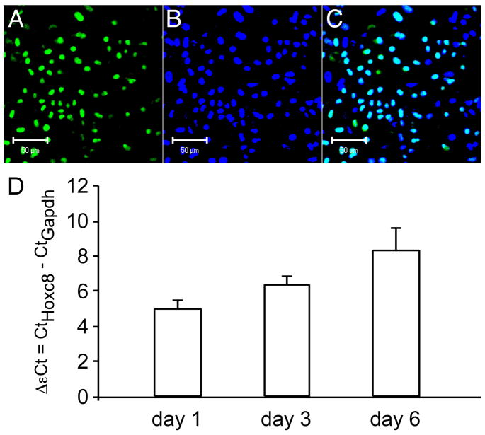
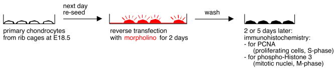
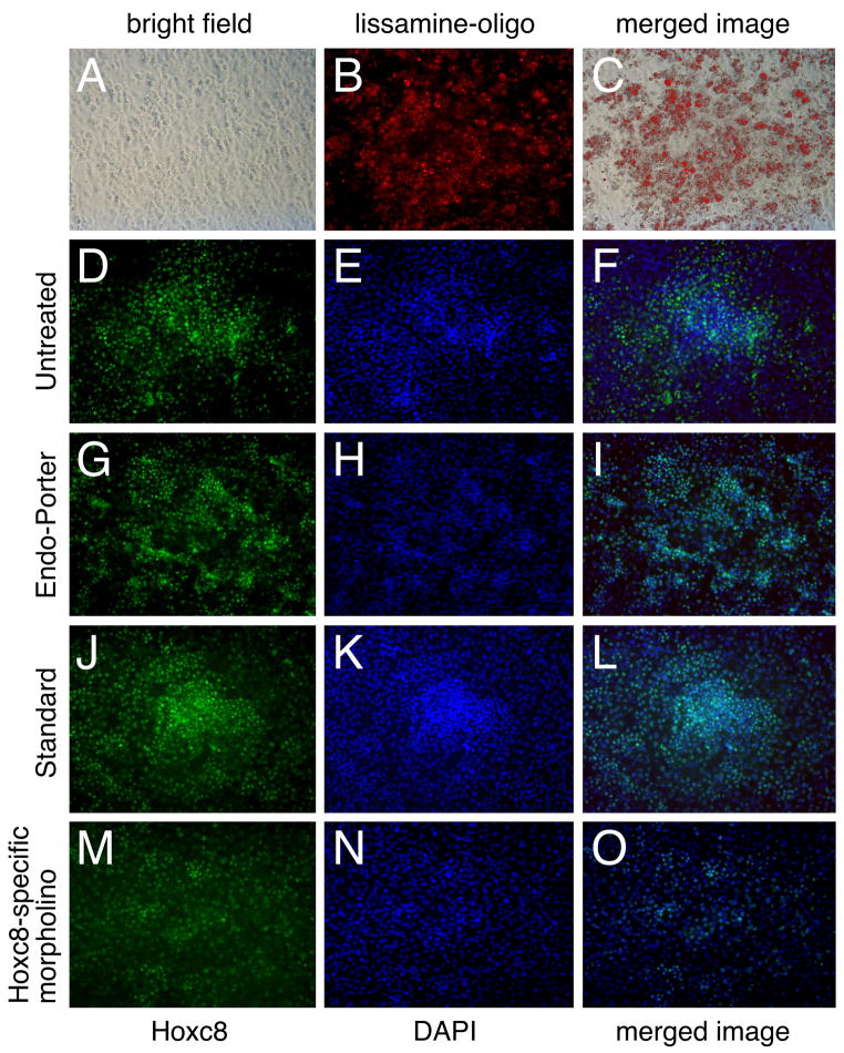
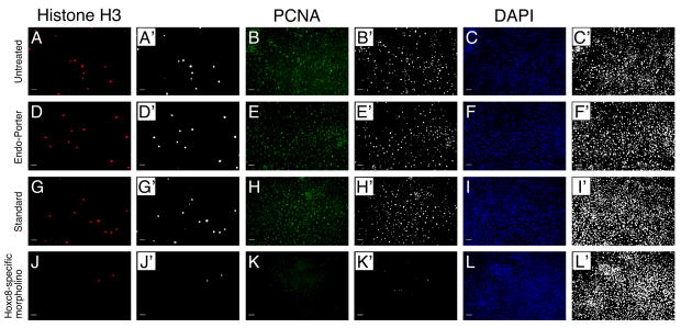
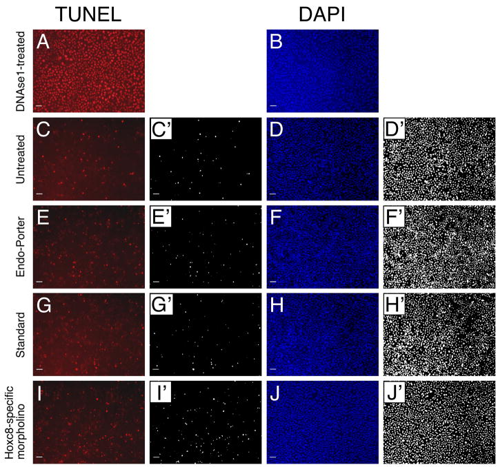
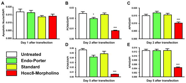
Numerous experiments in mutant and transgenic mice have implicated Hox transcription factors in development of the skeletal system, postulating a role for these proteins in cell proliferation of precursor cells and regulation of cell differentiation. Our own data from Hoxc8 and Hoxd4 transgenic mice suggest that Hoxc8 is involved in cell proliferation during cartilage development. In order to directly assess its role in cell proliferation of a specific skeletal cell type, the cartilage-producing chondrocyte, we performed morpholino-mediated knockdown experiments in normal primary chondrocytes. Through analysis of PCNA expression and staining for phosphorylated Histone 3, two cell cycle markers, we show that interference with Hoxc8 expression in chondrocytes reduces cell proliferation, but in the absence of apoptosis. Instead, cells with a knockdown in Hoxc8 expression appear to be delayed in their progression through the cell cycle. Our results provide evidence for prolonged duration of and delayed exit from M-phase, thus implicating a role for Hoxc8 in controlling cell cycle progression at this critical check point.


Figures

Figure 1
Figure 1. Expression of Hoxc8 in primary chondrocyte cultures
Immunolocalization of Hoxc8: Chondrocytes were fixed and immunostained for Hoxc8 on day 6 of culture. Slides were mounted with media containing DAPI and examined under a Leica confocal microscope. (A) Nuclear staining for Hoxc8; (B) DAPI staining of total cell nuclei; (C) Merged images for DAPI and Hoxc8. No signal was detected when the primary antibody was omitted. D) Quantitative RT-PCR for Hoxc8. The levels of Hoxc8 (as detected by cycle number above threshold in each sample, Ct) were normalized to the levels of the reference gene in the same sample and are expressed as ΔCt (ΔCt = CtHoxc8 − CtGapdh). Please note that lower bars (smaller ΔCt) reflect higher levels of expression.
Figure 2
Figure 2
Graphical representation of experimental approach.
Figure 3
Figure 3. Primary chondrocytes 2 days after reverse transfection with additional 2 days of culture
(A): Bright field picture of culture; (B): Lissamine-tagged morpholino oligos ingested by cells; (C): Merged image of bright field (A) and fluorescence image (B); (D–F) Untreated cultures; (G–I) Cultures to which only Endo-Porter was added; (J–L) Cultures transfected with irrelevant sequence morpholino (“standard”); (M–O) Cultures transfected with Hoxc8-specific morpholino. (D, G, J, M): Immunofluorescence for Hoxc8. (E, H, K, N): Nuclear staining with DAPI. (F, I, L, O): Merged images of Hoxc8 fluorescence and DAPI staining.
Figure 4
Figure 4. Morpholino-mediated knockdown of Hoxc8 affects cell proliferation
Primary chondrocyte cultures stained with anti-phospho-histone H3 antibody (A, D, G, J), PCNA-specific antibody (B, E, H, K) and for DAPI (C, F, I, L). (A–C): Untreated cultures (D–F): Cultures incubated with Endo-Porter alone. (G–I): Cultures transfected with 10μM standard oligo. (J–L): Cultures transfected with 10μM Hoxc8-specific morpholino oligo. The scale bar represents 100μM. All images labeled by apostrophes were converted to black and white format using the same threshold for quantification of pixel number (see Methods).
Figure 5
Figure 5. Minimal cell death in morpholino-exposed primary mouse chondrocyte cultures
(A, B) DNAseI-treated chondrocytes as positive control for TUNEL staining (C, D) Untreated chondrocytes (E, F) Endo-Porter-exposed chondrocytes (G, H) Chondrocytes transfected with standard oligo nucleotide (I, J) Chondrocytes transfected with Hoxc8-specific morpholino Panels labeled with apostrophe depict results used for quantification (see Methods).
Figure 6
Figure 6. Knockdown of Hoxc8 in reduces cell proliferation and delays cell cycle progression
(A): Chondrocyte cultures transfected with Hoxc8-specific morpholino show no difference in the apoptotic cell number compared to untreated and control groups on day 1 after transfection. (B – E): Time-course of chondrocyte proliferation after morpholino transfection. (B, D): Transfection with Hoxc8 specific antisense oligos reduced the number of cells entering S phase more than 70% on 2 days and 5 days after transfections. Although compared to the number of cells entering the S phase in untreated cultures, there is a decrease of PCNA-positive cells treated with Endo-Porter alone on day 2, there is no significant difference between untreated cells and cells transfected with the standard oligo. On day 5 there is no significant difference in the number of cells entering S phase in any of the control conditions, but PCNA-positivity is significantly reduced with Hoxc8-specific morpholino transfection. (C, E): Transfection with Hoxc8-specific antisense oligos reduced the number of cells entering M phase to less than 60% of controls by day 2 after transfection, and to less than 30% of controls by day 5 after transfection. However, there is no significant difference between the number of cells entering the M phase in untreated culture compared to cells treated with Endo-Porter alone or with standard control (*P<0.05, ***P<0.001).

References

    1. Capecchi MR. Function of homeobox genes in skeletal development. Ann NY Acad Sci. 1996;785:34–37. - PubMed
    1. Kappen C, Neubuser A, Balling R, Finnell R. Molecular basis for skeletal variation: Insights from developmental genetic studies in mice. Birth Defects Res B Dev Reprod Toxicol. 2007;80:425–50. - PMC - PubMed
    1. Newman SA. Sticky fingers: Hox genes and cell adhesion in vertebrate limb development. Bioessays. 1996;18:171–74. - PubMed
    1. Yokouchi Y, Nakazato S, Yamamoto M, Goto Y, Kameda T, Iba H, Kuroiwa A. Misexpression of hoxa-13 induces cartilage homeotic transformation and changes cell adhesiveness in chick limb buds. Genes Develop. 1995;9:2509–22. - PubMed
    1. Duboule D. Vertebrate hox-genes and proliferation: An alternative pathway to homeosis? Curr Opin Genet Dev. 1995;5:525–28. - PubMed

Publication types