. 2009 Jan;9(1):287-91.
doi: 10.1021/nl802915q.

Engineering of hetero-functional gold nanorods for the in vivo molecular targeting of breast cancer cells

Affiliations

Engineering of hetero-functional gold nanorods for the in vivo molecular targeting of breast cancer cells

Mohammad Eghtedari et al. Nano Lett. 2009 Jan.

Abstract

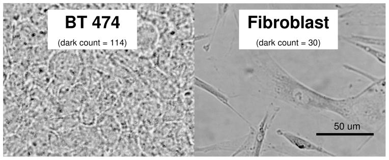
A novel technique is described to functionalize gold nanorods (GNRs) allowing for in vivo targeting of breast cancer tumors grown in athymic nude mice. GNRs were functionalized by covalent attachment of Herceptin (HER), a monoclonal antibody that enables molecular recognition of breast cancer cells expressing highly specific tumor associated antigens, and poly(ethylene glycol) (PEG) which obscures particles against the reticuloendothelial system in the body. The stability and functionality of fabricated particles (Her-PEG GNRs) were demonstrated in vitro in the presence of blood and then in vivo in nude mice model for breast cancer. The results demonstrate successful tumor accumulation of functionalized gold nanorods within HER2/neu overexpressing breast tumors in tumor-bearing nude mice and support the notions that GNRs can be used for molecular imaging of tumor.


Figures

Figure 1
Figure 1
Schematics of Her-PEG GNRs. The particles are attached covalently to Herceptin through Nanothinks Acid. PEG-Thiol molecules are attached to gold nanorods through their thiol functional group.
Figure 2
Figure 2
Three types of GNRS were incubated with three cell lines in-vitro for 24 hours. The number of dead cells in each treatment group was counted after staining cells with Trypan Blue. CTAB-coated GNRs increased the number of dead cells in-vitro while PEG and Her-PEG GNRs did not increase the number of dead cells under similar conditions. CTL reflects cells that were not treated with gold nanoparticles (Control). Bars indicate standard deviation.
Figure 3
Figure 3
Bright field images of four cell lines that were incubated with Her-PEG GNRs in-vitro. Slides were stained using a sliver staining kit to visualize gold nanorods as dark spots under conventional bright field microscopy. A: MCF-7 (Breast cancer but no HER2/neu expression→ no staining), B: Fibroblast (no HER2/neu expression → no staining), C: SK-BR3 (over expression of HER2/neu), D: BT-474 (over expression of HER2/neu), E: MCF-10A (normal breast, no HER2/neu → no staining). Dark counts were determined manually using cell count plugins and Image J software.
Figure 4
Figure 4
Bright field microscopic images of SK-BR3 cells that were incubated with PEG GNRs (A) or Her-PEG GNRs (B), Her GNRs (C), or no GNRs (D) in-vitro. Samples were silver-stained to reveal GNR as dark spots. The total number of dark spots, that indicate GNRs, is higher when Her-PEG GNRs or Her GNRs were used as compared to the groups were PEG GNRs or no GNRs were used.
Figure 5
Figure 5
Absorption spectra of supernatant solution after incubating three different types of GNRs for three hours in mouse heparinized blood. The disappearance of the peak absorption around 760 nm in the CTAB-Coated GNRs following incubation in blood indicated the aggregation of CTAB-coated GNRs in the blood while the presence of a preserved peak absorption at 760 nm for particles functionalized with the PEG GNRs and Her-PEG GNRs following incubation in blood confirm the stability of these particles within blood microenvironment.
Figure 6
Figure 6
Bright field images of BT-474 and Fibroblast cells incubated with Her-PEG GNRs that were premixed with mouse blood for four hours. Samples were stained with silver enhancement kit and examined under bright field microscopy to reveal targeted GNRs. Dark counts were determined on each image manually using image J software.
Figure 7
Figure 7
Typical bright field images of mouse tissue slices following intravenous injection of GNRs to ten nude mice. 100 μL of PBS, PEG GNRs, or Her-PEG GNRs was injected intravenously into the tail vein and animals were sacrificed 24 hours post injection. Samples of tumor, liver, and spleen were obtained and silver stained to reveal gold nanorods. The images in the two bottom rows exhibit darker spots caused by enhanced accumulation of GNRs.

References

    1. Ferrari M. Nature Reviews Cancer. 2005;5(3):161–171. - PubMed
    1. Niidome T, Yamagata M, Okamoto Y, Akiyama Y, Takahashi H, Kawano T, Katayama Y, Niidome Y. Journal of Controlled Release. 2006;114(3):343–347. - PubMed
    1. Connor EE, Mwamuka J, Gole A, Murphy CJ, Wyatt MD. Small. 2005;1(3):325–327. - PubMed
    1. Ditrich H, Splechtna H. Tissue & Cell. 1988;20(6):891–898. - PubMed
    1. O’Neal DP, Hirsch LR, Halas NJ, Payne JD, West JL. Cancer Letters. 2004;209(2):171–176. - PubMed

Publication types