Review
. 2009 Jun;11(6):1313-33.
doi: 10.1089/ars.2008.2363.

Signaling components of redox active endosomes: the redoxosomes

Affiliations
Review

Signaling components of redox active endosomes: the redoxosomes

Fredrick D Oakley et al. Antioxid Redox Signal. 2009 Jun.

Abstract

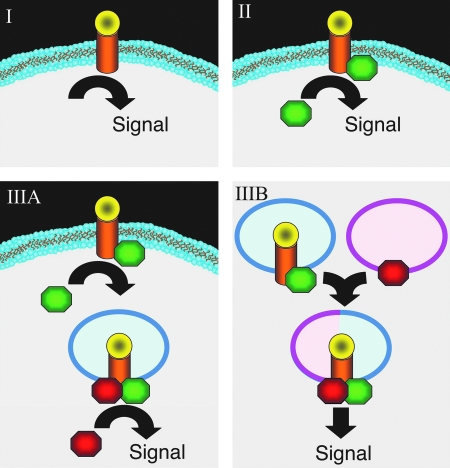
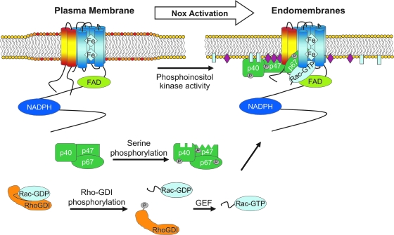
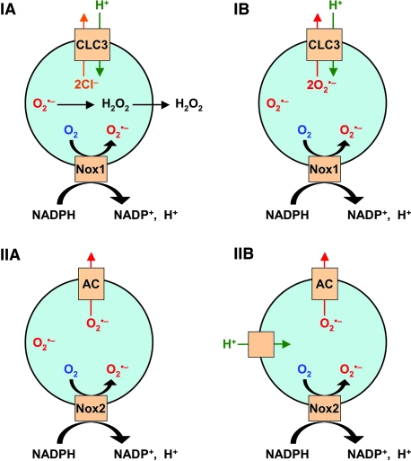
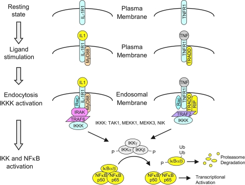
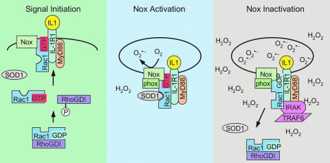
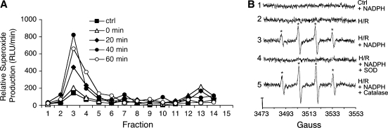
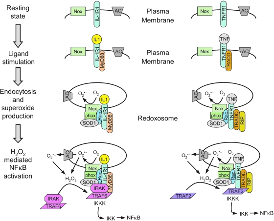
Subcellular compartmentalization of reactive oxygen species (ROS) plays a critical role in transmitting cell signals in response to environmental stimuli. In this regard, signals at the plasma membrane have been shown to trigger NADPH oxidase-dependent ROS production within the endosomal compartment and this step can be required for redox-dependent signal transduction. Unique features of redox-active signaling endosomes can include NADPH oxidase complex components (Nox1, Noxo1, Noxa1, Nox2, p47phox, p67phox, and/or Rac1), ROS processing enzymes (SOD1 and/or peroxiredoxins), chloride channels capable of mediating superoxide transport and/or membrane gradients required for Nox activity, and novel redox-dependent sensors that control Nox activity. This review will discuss the cytokine and growth factor receptors that likely mediate signaling through redox-active endosomes, and the common mechanisms whereby they act. Additionally, the review will cover ligand-independent environmental injuries, such as hypoxia/reoxygenation injury, that also appear to facilitate cell signaling through NADPH oxidase at the level of the endosome. We suggest that redox-active endosomes encompass a subset of signaling endosomes that we have termed redoxosomes. Redoxosomes are uniquely equipped with redox-processing proteins capable of transmitting ROS signals from the endosome interior to redox-sensitive effectors on the endosomal surface. In this manner, redoxosomes can control redox-dependent effector functions through the spatial and temporal regulation of ROS as second messengers.


Figures

FIG. 1.
FIG. 1.
Mechanisms of receptor signaling following ligand binding at the plasma membrane. (I) Ligand binding directly activates a receptor-mediated cell signaling event at the plasma membrane in the absence of additional cytosolic effectors. (II) Ligand binding leads to the recruitment of a cytosolic effector to the receptor at the plasma membrane, which in turn initiates signaling. (IIIA) Ligand binding and cytosolic effector recruitment initiates endocytosis of the receptor. Following endocytosis and the formation of a specific microenvironment, downstream effectors are recruited to drive formation of an active signaling complex. (IIIB) Ligand binding and cytosolic effector recruitment initiate endocytosis of the receptor, as shown in IIIA. However, the formation of a signaling complex requires endosomal fusion with another cellular compartment to generate the microenvironment needed for receptor complex activation. Such microenvironmental changes may include the docking of secondary membrane associated effectors (as shown) and/or changes in endosomal membrane structure/function (i.e., the composition of phospholipid or ion channels or endosomal pH). (For interpretation of the references to color in this figure legend, the reader is referred to the web version of this article at www.liebertonline.com/ars).
FIG. 2.
FIG. 2.
Application of BSA-H2HFF fluorescence to visualize redoxosomal ROS following stimulation by TNFα or IL-1β. (A, B) MCF-7 cells were stimulated for 20 min in the presence or absence of 5 ng/ml IL-1β and BSA-H2HFF, as marked. (C, D) A vascular smooth muscle cell line was stimulated for 20 min in the presence or absence of 10 ng/ml TNFα and BSA-H2HFF, as marked. Nuclei are marked by DAPI (blue) and oxidized H2HFF fluorescence is in green. (For interpretation of the references to color in this figure legend, the reader is referred to the web version of this article at www.liebertonline.com/ars).
FIG. 3.
FIG. 3.
Cytochemical detection of redoxosomal superoxides at the ultrastructural EM level, using a 3,3′-diaminobenzidine (DAB)-Mn2+ reaction. MCF-7 cells were stimulated with 0.5 ng/ml IL-1β for 15 min, followed by staining in Hepes Buffer (10 mM Hepes, pH 7.4, 135 mM NaCl, 5 mM KCl, 1 mM CaCl2) containing 5 mM DAB, 1 mM MnCl2, and 1 mM NaN3 for 5 min at 37°C. After washing with Hepes buffer, cell samples were fixed for electron microscopy using 2% glutaraldehyde at 4°C overnight, and washed with Hepes buffer three times. Post-fixation was performed with 1% osmium tetroxide for 1 h, followed by washing three times in buffer alone. Cells were then dehydrated through a graded series of ethanol solutions (from 50%, 75%, and 95%, to 100%) and embedded in Epon resin. Sections (100 nm) were then evaluated by TEM. (AD) MCF-7 cells were treated with (A) PBS or (BD) IL-1β for 15 min and stained in the presence of DAB/Mn2+ for 5 min, followed by fixation and evaluation by TEM. Two endosomal populations were seen, including those with electron-dense precipitates in their interiors (solid arrows) and those lacking a precipitate (open arrows). An enlargement of the boxed region in (C) is shown in (D). Mitochondria are marked by an “m” for reference. Arrows mark regions enlarged in the inset.
FIG. 4.
FIG. 4.
Model for Nox2 activation during redoxosomal signaling. In the resting state, Nox2 (protein with six blue transmembrane domains) and p22phox (protein with two yellow/red transmembrane domains) make up the flavocytochrome b558 complex, which is located in lipid rafts of the plasma membrane (marked by a widened lipid domain). The Nox subunits p40phox, p47phox, and p67phox are found in a complex in the cytosol, and Rac is in its inactive GDP-bound state associated with RhoGDI. In response to a stimulus, RhoGDI is phosphorylated, causing it to disassociate from Rac-GDP, which exposes its prenylated tail. Prenylated Rac-GDP subsequently interacts with a GEF that exchanges GDP for GTP, generating an activated Rac-GTP complex. Rac1 then moves to the membrane, where it interacts with Nox2. Phosphorylation of p40phox, p47phox, and/or p67phox recruits these subunits to the Nox complex. The serine-phosphorylated p47phox is stabilized on the Nox complex, by interacting with both p22phox (via its N-terminal SH3 domain) and PI(3,4)P2 (via its PX domain). p40phox may further stabilize the complex along the membrane by interacting with PI(3)P via its PX domain. p67phox binds to Nox2 via its activation domain, and also associates with Rac-GTP via its N-terminal tetratricopeptide repeats. Following the recruitment of all members of the Nox complex, superoxide production takes place as NADPH binds to the cytoplasmic tail of Nox2. Electrons are transferred to FAD, which is also bound to the Nox2 cytoplasmic tail. FAD then sequentially transfers the electrons to the Nox complex, where they pass through the two heme centers, and are ultimately transferred to oxygen on the luminal side of the redoxosome. (For interpretation of the references to color in this figure legend, the reader is referred to the web version of this article at www.liebertonline.com/ars).
FIG. 5.
FIG. 5.
Possible mechanisms of charge neutralization and ROS production by redoxosomes. (IA) The mechanism proposed by Miller et al. (78) for Nox1 redoxosomes in IL-1β-stimulated aortic smooth muscle cells. In this model, the voltage gradient is neutralized by the anion channel ClC-3, which acts as a proton-chloride antiporter. ROS signaling relies on spontaneous dismutation of superoxide to hydrogen peroxide within the redoxosome lumen, followed by diffusion of hydrogen peroxide into the cytosol for redox signaling. (IB) In an open alternative possibility to the Miller model, ClC-3 acts as a proton-superoxide antiporter, and thereby accomplishes charge neutralization and the transport of superoxide into the cytosol for redox signaling. (IIA) A proposed model for IL-1β-stimulated Nox2 redoxosome charge neutralization and ROS production by MCF-7 mammary epithelial cells (83). In this model, an anion channel neutralizes charge by transporting superoxide out of the redoxosome. (IIB) In an extension of model IIA, a NFA/DIDS-insensitive proton channel compensates for changes in lumenal pH produced by Nox2. (For interpretation of the references to color in this figure legend, the reader is referred to the web version of this article at www.liebertonline.com/ars).
FIG. 6.
FIG. 6.
Relevant adaptors and effectors of the IL-1R1 and TNFR1 signaling pathways that lead to NFκB activation. Receptors are drawn as monomers for simplicity of presentation, but actually occur as dimers of IL-1R1 and IL-1 receptor accessory protein (IL-1RAcP), and tetramers of TNFR1, in their ligand-bound states. The molar ratios of adaptor/effectors are also not drawn for accuracy, but for clarity of the types of molecular interactions. Potential IKK kinases (IKKKs) that phosphorylate the IKK complex are listed. Not all potential adaptors of these receptor pathways are listed, but we have attempted to include those most relevant to redoxosomal signaling. (For interpretation of the references to color in this figure legend, the reader is referred to the web version of this article at www.liebertonline.com/ars).
FIG. 7.
FIG. 7.
Redox-sensor model for controlling Nox activation in redoxosomes. The proposed regulatory model for IL-1β signaling builds on the finding that SOD1 is actively recruited to IL-1β-stimulated redoxosomes, and binds to Rac1 in a redox-dependent fashion to control Rac1 GTPase activity. Left panel: In the resting state, Rac1-GDP remains inactive because it is bound to RhoGDI. In this state the majority of Rac1 is most likely in a reduced state, due to the reducing conditions in the cytoplasm. Following IL-1β stimulation, RhoGDI is phosphorylated, releasing Rac-GDP into the cytosol. A Rac1-GEF then activates Rac1 by exchanging GDP for GTP, allowing for the association of Rac1-GTP with the Nox complex at the plasma membrane. IL-1β binding to IL-1R1 also leads to docking of MyD88 at the plasma membrane, which initiates endocytosis of the receptor/Nox complex. Center panel: Following or during early stages of endocytosis, SOD1 binds to reduced Rac1-GTP associated with the Nox complex, maintaining Rac1 in its active GTP-bound conformation by inhibiting the GTPase activity of Rac1. Superoxide production by redoxosomes is initiated at this stage, and Nox activity is maximal. Right panel: The localized buildup of superoxide and hydrogen peroxide around the redoxosome creates an oxidative microenvironment that promotes IRAK/TRAF6 complex docking on the receptor. Eventually, the increased levels of hydrogen peroxide oxidize Rac1, resulting in the disassociation of SOD1 from Rac-GTP, and hydrolysis of GTP by Rac1. Nox activity is terminated as Rac-GDP is formed on the redoxosome and Rac1 is recycled from the membrane by RhoGDI. (For interpretation of the references to color in this figure legend, the reader is referred to the web version of this article at www.liebertonline.com/ars).
FIG. 8.
FIG. 8.
H/R induction of NADPH-dependent superoxide production by the endosomal compartment. (A) TIB73 cells (a hepatocyte cell line) were treated in a hypoxic atmosphere for 5 h, and were then reoxygenated for various periods prior to subcellular fractionation by iodixanol gradient separation of postnuclear supernatants. For each fraction, relative NADPH-dependent superoxide production was detected by lucigenin luminescence, and is shown as Relative Light Units (RLU) per minute. Control (ctrl) cells were not subjected to H/R. H/R experimental samples are shown at times post-reoxygenation (0–60 min). The zero minute post-reoxygenation time point underwent hypoxia for 5 h, but did not undergo reoxygenation. (B) Superoxide production in the peak vesicular fractions (#3-4) of normoxic (ctrl) and 40-min post-reoxygenation TIB73 cell samples was evaluated by ESR, using the DMPO spin-trap in the presence or absence of 100 μM of NADPH to stimulate Nox activity (conditions 1–3). Assays were also performed with isolated vesicular fractions pre-loaded with SOD1 or catalase proteins, by adding 1 mg/ml to the reoxygenation media (conditions 4 and 5). Asterisks mark DMPO/OH adducts derived from superoxides (as demonstrated by SOD1, but not catalase, quenching following loading of the endosomal lumen). The y-axis represents 5 × 104 arbitrary units of intensity, and the x-axis represents the magnetic field in Gauss.
FIG. 9.
FIG. 9.
Model of redoxosomal signaling for IL-1β and TNFα pathways. In the resting state, the cell expresses Nox, an anion channel(s) (AC), and IL-1R1 or TNFR1 on the plasma membrane. Following receptor binding by IL-1β or TNFα, pathway-specific early effectors (MyD88 or TRADD) are recruited to their cognate receptor at the plasma membrane and initiate endocytosis. Rac1 recruits Nox into the early endosome by tethering (indirectly or directly) the receptor to the Nox complex. Phox subunits (p47phox and p67phox in the case of Nox2) are recruited to the newly formed endosomes to produce superoxide-generating redoxosomes. It is currently unclear at which point SOD1 is recruited to Rac1 on redoxosomes. However, this process appears to be important for maintaining Rac1 in a GTP-bound active state. Membrane-impermeable superoxide generated in the redoxosome lumen may spontaneously dismutate to hydrogen peroxide and pass through the endosomal membrane and out of the redoxosome. Alternatively, a DIDS/NFA-sensitive chloride channel may transport the superoxide out of the redoxosome, where it is subsequently converted to hydrogen peroxide. The localized production of hydrogen peroxide at the redoxosome surface leads to transmission of redox-specific signals to either the receptor or one of the downstream effectors (IRAK/TRAF6 or TRAF2), allowing for docking of these TRAF effectors and subsequent activation of pathway-specific IKKKs. These IKKKs in turn phosphorylate the IKK complex, which leads to NFκB activation. The redoxosomal-signaling pathway is downregulated as the build up of cytosolic hydrogen peroxide oxidizes Rac1 on the surface of the endosome, and this causes disassociation of SOD1 from Rac1 (not shown). In the absence of SOD1 binding to Rac1, Rac1 quickly hydrolyzes GTP and becomes inactive, leading to the termination of Nox-dependent superoxide production. (For interpretation of the references to color in this figure legend, the reader is referred to the web version of this article at www.liebertonline.com/ars).

References

    1. Abe J. Takahashi M. Ishida M. Lee JD. Berk BC. c-Src is required for oxidative stress-mediated activation of big mitogen-activated protein kinase 1. J Biol Chem. 1997;272:20389–20394. - PubMed
    1. Ando S. Kaibuchi K. Sasaki T. Hiraoka K. Nishiyama T. Mizuno T. Asada M. Nunoi H. Matsuda I. Matsuura Y, et al. Post-translational processing of rac p21s is important both for their interaction with the GDP/GTP exchange proteins and for their activation of NADPH oxidase. J Biol Chem. 1992;267:25709–25713. - PubMed
    1. Arcaro A. Aubert M. Espinosa del Hierro ME. Khanzada UK. Angelidou S. Tetley TD. Bittermann AG. Frame MC. Seckl MJ. Critical role for lipid raft-associated Src kinases in activation of PI3K-Akt signalling. Cell Signal. 2007;19:1081–1092. - PubMed
    1. Babior BM. Kipnes RS. Curnutte JT. Biological defense mechanisms. The production by leukocytes of superoxide, a potential bactericidal agent. J Clin Invest. 1973;52:741–744. - PMC - PubMed
    1. Banfi B. Clark RA. Steger K. Krause KH. Two novel proteins activate superoxide generation by the NADPH oxidase NOX1. J Biol Chem. 2003;278:3510–3513. - PubMed

Publication types