. 2009 Mar;11(3):425-38.
doi: 10.1089/ars.2008.2235.

Sensory neurons and schwann cells respond to oxidative stress by increasing antioxidant defense mechanisms

Affiliations

Sensory neurons and schwann cells respond to oxidative stress by increasing antioxidant defense mechanisms

Andrea M Vincent et al. Antioxid Redox Signal. 2009 Mar.

Abstract

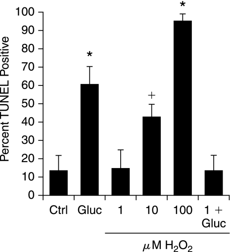
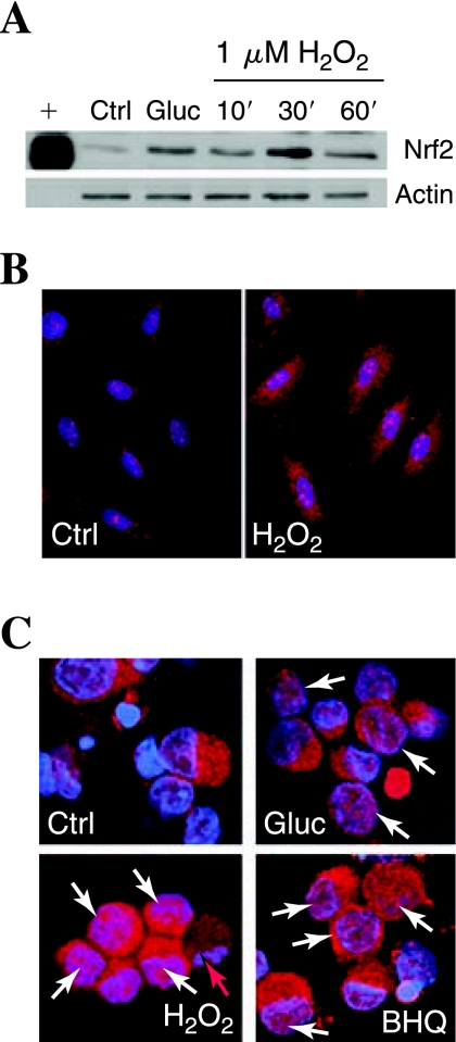
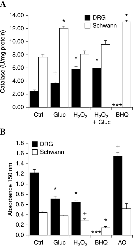
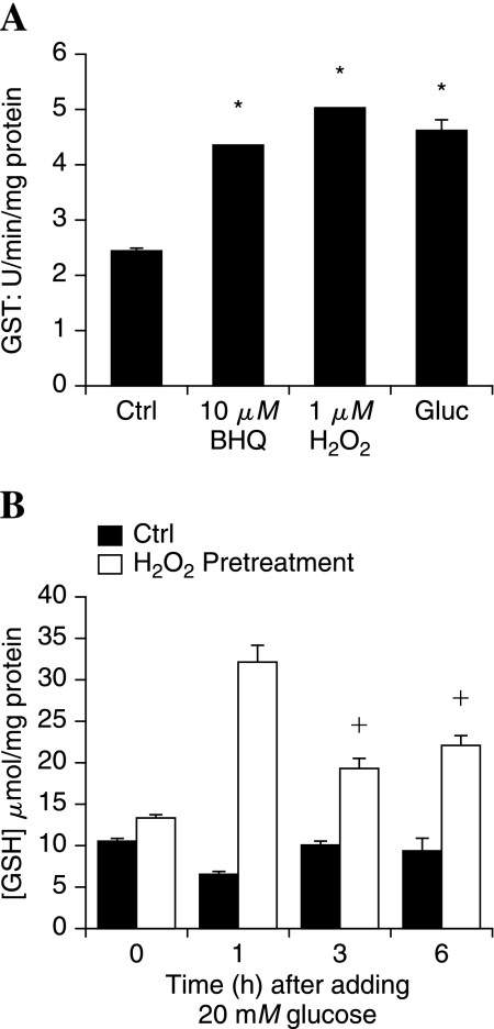
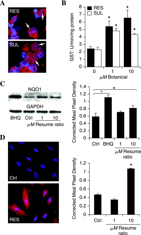
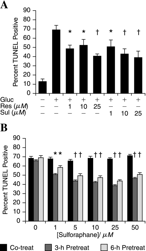
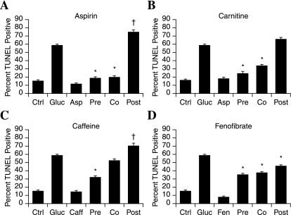
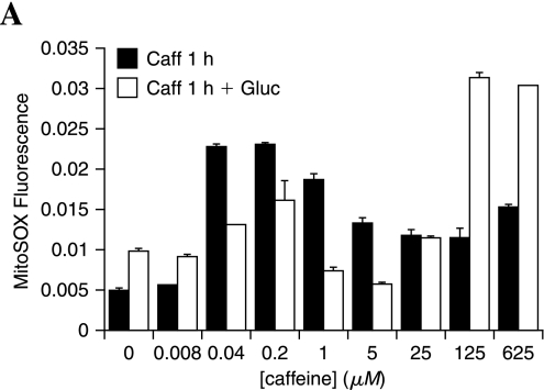
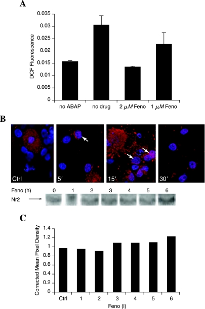
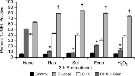
Elevated blood glucose is a key initiator of mechanisms leading to diabetic neuropathy. Increases in glucose induce acute mitochondrial oxidative stress in dorsal root ganglion (DRG) neurons, the sensory neurons normally affected in diabetic neuropathy, whereas Schwann cells are largely unaffected. We propose that activation of an antioxidant response in DRG neurons would prevent glucose-induced injury. In this study, mild oxidative stress (1 microM H2O2) leads to the activation of the transcription factor Nrf2 and expression of antioxidant (phase II) enzymes. DRG neurons are thus protected from subsequent hyperglycemia-induced injury, as determined by activation of caspase 3 and the TUNEL assay. Schwann cells display high basal antioxidant enzyme expression and respond to hyperglycemia and mild oxidative stress via further increases in these enzymes. The botanical compounds resveratrol and sulforaphane activate the antioxidant response in DRG neurons. Other drugs that protect DRG neurons and block mitochondrial superoxide, identified in a compound screen, have differential ability to activate the antioxidant response. Multiple cellular targets exist for the prevention of hyperglycemic oxidative stress in DRG neurons, and these form the basis for new therapeutic strategies against diabetic neuropathy.


Figures

FIG. 1.
FIG. 1.
Mild prooxidant stress prevents dorsal root ganglion (DRG) neuron injury in subsequent exposure to hyperglycemia. DRG neurons were exposed to a range of concentrations of H2O2 (1–100 μM) and then examined for cell injury by TUNEL after 24 h. Hyperglycemia was modeled by adding 20 mM glucose, giving a final concentration of 45 mM glucose (Gluc). In the last bar, DRG neurons were preincubated with the nontoxic concentration of H2O2 (1 μM) for 3 h before hyperglycemia. Values are expressed as mean ± SEM. *p < 0.01 versus control (Ctrl). +p < 0.05 versus Ctrl.
FIG. 2.
FIG. 2.
Prooxidants induce Nrf2 accumulation in the nucleus. (A) Expression of Nrf2 was assessed in Schwann cells with Western blotting. Actin was used as a loading control (Ctrl). Schwann cells were exposed to 20 mM added glucose (Gluc) or 1 μM H2O2 for 10, 30, or 60 min. A positive control (+) was compared with the primary cell samples. (B, C) The nuclear localization of Nrf2 was assessed by staining red with an antibody for Nrf2 and counterstaining the nuclei blue by using bis-benzimide. In all conditions shown, cells were fixed and stained after 30-min treatment. Colocalization of Nrf2 with nucleus appears pink and is indicated with white arrows. Representative images of Schwann cells are shown in (B), and dorsal root ganglion (DRG) neurons, in (C). (D) Red arrow, A DRG neuron undergoing programmed cell death, with condensed and fragmented chromatin.
FIG. 3.
FIG. 3.
Mild prooxidant stress activates antioxidant enzymes. Dorsal root ganglion (DRG) neurons or Schwann cells were exposed to hyperglycemia (20 mM added glucose), mild prooxidant [1 μM H2O2 or 10 μM tert-butylhydro-quinout (BHQ)], or the antioxidant (AO) α-lipoic acid (100 μM) for 3 h. H2O2 + glucose indicates 3-h H2O2 followed by 3-h glucose. After 3 h, antioxidant enzyme activities were measured: (A) Catalase, (B) HO-1. +p < 0.05 compared with control (Ctrl). *p < 0.01 compared with Ctrl.
FIG. 4.
FIG. 4.
Oxidant stress preconditions DRG neurons to maintain cellular GSH in the presence of hyperglycemia. (A) DRG neurons were treated with mild prooxidants or 20 mM added glucose (Gluc), and then the activity of GST was measured in cell lysates after 3 h. *p < 0.01 compared with untreated control (Ctrl). (B) DRG neurons were pretreated for 3 h with H2O2 (1 μM) and then exposed to 20 mM added glucose for 0, 1, 3, or 6 h. DRG neurons were lysed, and the concentration of reduced glutathione (GSH) was measured. +p < 0.05 compared with no pretreatment (Ctrl). *p < 0.01 compared with no pretreatment.
FIG. 5.
FIG. 5.
Botanical compounds activate Nrf2 translocation and the antioxidant response. DRG neurons were exposed to 1 or 10 μM resveratrol (RES) or sulforaphane (SUL), and then the antioxidant response was assessed. (A) 1 h after exposure to 10 μM compound, the neurons were fixed and labeled for Nrf2 (red) or nuclei (bis-benzimide, blue). Nuclear Nrf2 is indicated with white arrows. (B) The activity of GST was determined after 3 h. *p < 0.05 compared with control (0 μM). (C) The expression of NQO1 protein was determined with Western blotting after 3-h RES. The prooxidant BHQ (10 μM) was included for comparison. Pixel density of the NQO1 band in each condition was normalized to the corresponding GAPDH band. (D) Schwann cell antioxidant response to resveratrol (10 μM) was assessed. Nrf2 localization after 1 h (left) and NQO1 expression after 3 h (right). Corrected mean pixel density represents the mean ± SEM for each condition from three replicate blots. †p < 0.01 and *p < 0.05 versus untreated control (C).
FIG. 6.
FIG. 6.
Resveratrol and sulforaphane pretreatment can prevent hyperglycemic injury in DRG neurons. (A) Increasing concentrations of resveratrol (Res) or sulforaphane (Sul) were applied to DRG neurons 3 h before the application of 20 mM glucose (Gluc). (B) Increasing concentrations of sulforaphane were applied to DRG neurons either at the same time (Co-treat), 3 h before, or 6 h before 20 mM added glucose. (A, B) Cells were fixed after 24 h and TUNEL stained. The bars represent the mean percentage TUNEL positive for three independent experiments ± SEM. †p < 0.01 and *p < 0.05 versus glucose only (A) or no pretreatment (B).
FIG. 7.
FIG. 7.
Compounds that prevent glucose-induced oxidative injury operate via different mechanisms. Four lead compounds that prevent DRG neuron mitochondrial oxidative stress and injury in hyperglycemia were assessed in different treatment paradigms in adult rat DRG neurons. The 1 μM compound was added at the same time as (Co), 3 h before (Pre), or 1 h after (Post) 20 mM glucose. The bars represent the mean percentage of TUNEL positive for two independent experiments ± SEM. *p < 0.01 compared with glucose only. +p < 0.05 compared with glucose only.
FIG. 8.
FIG. 8.
Caffeine promotes mitochondrial oxidative stress. Adult DRG neurons were exposed to increasing concentrations of caffeine alone for 1 h or caffeine for 1 h followed by 20 mM added glucose for 1 h. DRG neurons were loaded with MitoSOX and read at 485 nm ex, 590 em on a plate reader. Bars indicate the mean of three replicates + SEM.
FIG. 9.
FIG. 9.
Fenofibrate is an antioxidant that also activates Nrf2. (A) A cell-free assay was performed by mixing fenofibrate with DCFDA (10 μM) in HBSS solution, and then adding 50 mM oxidizing agent 2,2′-azobis (amidinopropane) dihydrochloride (ABAP). The oxidation of DCFDA to DCF was determined by measuring DCF fluorescence after 2 min in a plate reader. A control containing BSA instead of fenofibrate (no drug) was included for comparison. (B) The localization of Nrf2 was assessed with immunohistochemistry in adult DRG neuron cultures after exposure to 10 μM fenofibrate. Nrf2 was stained red, and the nuclei were counterstained blue by using bis-benzimide. Colocalization appears pink (white arrows). (C) Changes in Nrf2 expression were assessed with Western blotting after 1- to 6-h exposure to 1 μM fenofibrate. The representative blot with corresponding densitometry (normalized against GAPDH) suggests a modest increase in Nrf2 expression after 3 h, with a further increase by 6 h.
FIG. 10.
FIG. 10.
Differential ability of compounds of interest to activate the antioxidant response in DRG neurons. (A) Adult DRG neurons were exposed to 1 μM compounds or H2O2 for 3 h, and then Western blotted for NQO1. The bar graph illustrates the mean and SEM for three replicates normalized to actin, and below is a representative blot. *p < 0.05 compared with untreated control. The compounds are fenofibrate (Feno), caffeine (Caff), carnitine (Carn), and aspirin (Asp). (B, C) Adult DRG neurons were exposed to 1 μM fenofibrate for 1–6 h, and then Western blotted for NQO1 (B) or assayed for GST activity (C). *p < 0.05 compared with untreated control.
FIG. 11.
FIG. 11.
DRG neuron protection via Nrf2-activating drugs requires protein synthesis. Adult DRG neurons were exposed to 1 μM resveratrol (Res), sulforaphane (Sul), fenofibrate (Feno), or H2O2 for 3 h in the presence or absence of 20 μg/ml CHX. Glucose (20 mM) was then added, and cells were fixed after 24 h and TUNEL stained. *Drugs decreased DRG neuron injury compared with glucose only, p < 0.01. †CHX increased glucose-induced injury in the presence of the drugs, p < 0.05.

References

    1. Reducing ocular damage in type 2 diabetes: the FIELD study shows fenofibrate benefits. Cardiovasc J Afr. 2007;18:400. - PubMed
    1. Afaq F. Mukhtar H. Botanical antioxidants in the prevention of photocarcinogenesis and photoaging. Exp Dermatol. 2006;15:678–684. - PubMed
    1. Ahlgren-Beckendorf JA. Reising AM. Schander MA. Herdler JW. Johnson JA. Coordinate regulation of NAD(P)H:quinone oxidoreductase and glutathione-S-transferases in primary cultures of rat neurons and glia: role of the antioxidant/electrophile responsive element. Glia. 1999;25:131–142. - PubMed
    1. Almhanna K. Wilkins PL. Bavis JR. Harwalkar S. Berti-Mattera LN. Hyperglycemia triggers abnormal signaling and proliferative responses in Schwann cells. Neurochem Res. 2002;27:1341–1347. - PubMed
    1. Bonnard C. Durand A. Peyrol S. Chanseaume E. Chauvin MA. Morio B. Vidal H. Rieusset J. Mitochondrial dysfunction results from oxidative stress in the skeletal muscle of diet-induced insulin-resistant mice. J Clin Invest. 2008;118:789–800. - PMC - PubMed

Publication types