Review
. 2008 Mar;4(2):151-62.
doi: 10.2217/17455057.4.2.151.

Progesterone and breast cancer

Affiliations
Review

Progesterone and breast cancer

Carol A Lange et al. Womens Health (Lond). 2008 Mar.

Abstract

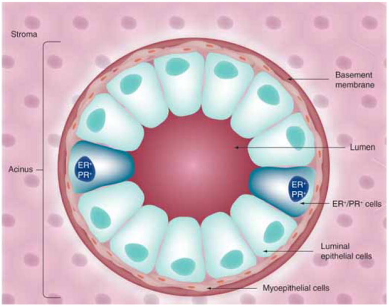
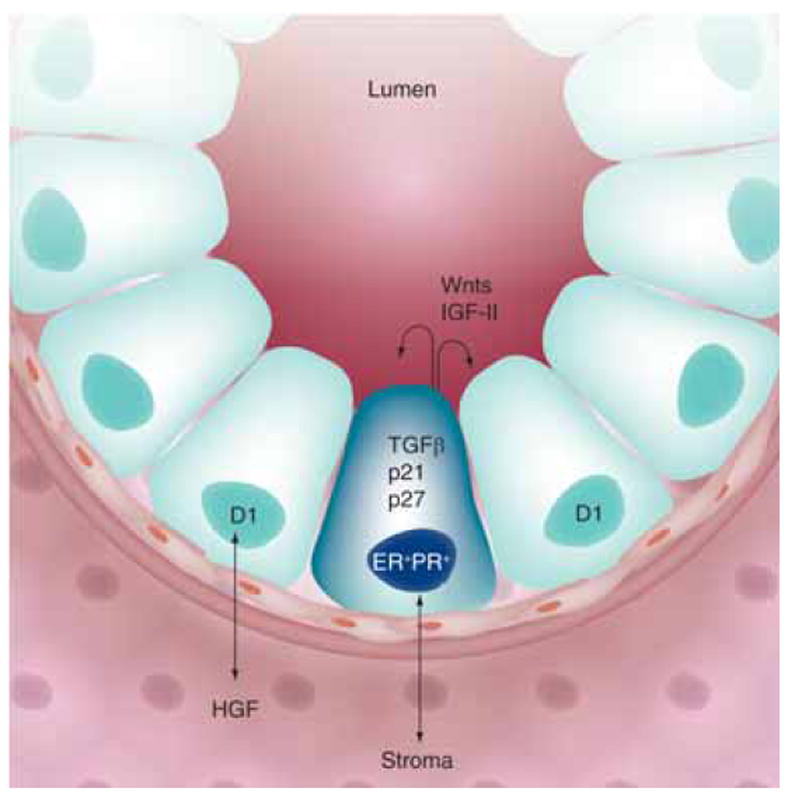
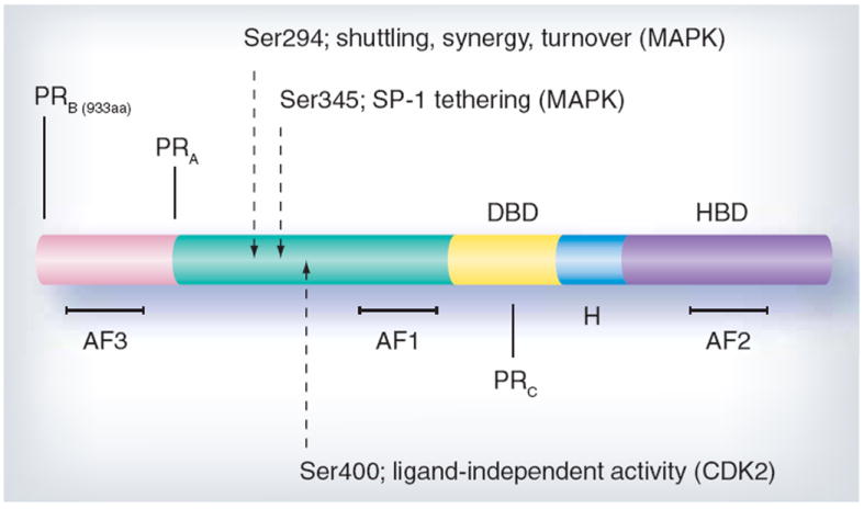
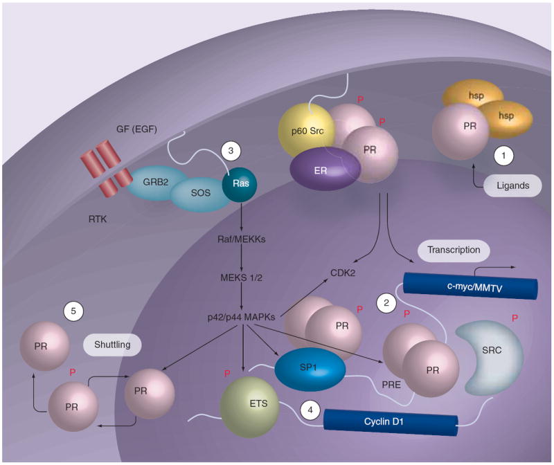
Progesterone is an ovarian steroid hormone that is essential for normal breast development during puberty and in preparation for lactation and breastfeeding. The actions of progesterone are primarily mediated by its high-affinity receptors, which include the classical progesterone receptor (PR)-A and -B isoforms, located in diverse tissues, including the brain, where progesterone controls reproductive behavior, and the breast and reproductive organs. Progestins are frequently prescribed for contraception or during postmenopausal hormone replacement therapy, in which progestins are combined with estrogen as a means to block estrogen-induced endometrial growth. The role of estrogen as a potent breast mitogen is undisputed, and inhibitors of the estrogen receptor and estrogen-producing enzymes (aromatases) are effective first-line cancer therapies. However, PR action in breast cancer is grossly understudied and remains controversial. Herein, we review existing evidence and discuss the challenges to defining a role for progesterone in breast cancer.


Conflict of interest statement

Financial & competing interests disclosure

The authors have no relevant affiliations or financial involvement with any organization or entity with a financial interest in or financial conflict with the subject matter or materials discussed in the manuscript.

No writing assistance was utilized in the production of this manuscript.

Figures

Figure 1
Figure 1. Mammary gland architecture
Acini, located at the ends of ducts in the mature mammary gland, are the functional units of the lactating mammary gland. Luminal epithelial cells (apical) are arranged as polar cells in contact with myoepithelial cells (basal). Epithelial cell populations are separated from the stromal environment by a basement membrane.
Figure 2
Figure 2. Paracrine signaling in the mammary gland
A minority population of steroid hormone receptor positive (ER+/PR+) cells occur adjacent to proliferating cells in the normal mammary gland. Communication between the epithelial and stromal compartments mediates proliferative paracrine signaling. Early events during breast cancer development may mediate switching from paracrine to autocrine mechanisms of proliferation.
Figure 3
Figure 3. Progesterone receptor structure
Progesterone receptor (PR)-B and -A are ligand-activated transcription factors containing a hormone-binding domain (HBD), hinge region (H), DNA-binding domain (DBD) and amino terminus. Activation functions (AFs) are the sites of coregulator interaction required for transcription. Serines 294, 345 and 400 are examples of regulatory sites that are phosphorlyated in response to progestins and/or mitogenic signaling pathways that modify PR function, in part by direct phosphorylation of PR or its coregulatory proteins.
Figure 4
Figure 4. Progesterone receptor function
Phosphorylation (P) of specific sites in PR couples multiple receptor functions, including transcriptional synergy in the presence of steroid hormones and growth factors predicted to activate MAPK and/or CDK2, and nuclear import or export (shuttling) in response to MAPK activation. Rapid ligand-dependent PR downregulation occurs by the ubiquitin–proteasome pathway (degradation). 1. Ligand-binding mediates dissociation of heat-shock proteins and nuclear accumulation of PR. 2. Nuclear PR mediates gene regulation via the classical pathway; phosphorylated PR may recruit regulatory molecules that are phosphoproteins, and function in one or more interconnected processes (transcription, localization and turnover). 3. PR and growth factors activate MAPKs independently via a c-Src kinase-dependent pathway, and this may result in positive regulation of PR action via ‘feedback’ regulation (i.e., direct phosphorylation of liganded PR or co-activators), occurring in both the absence and presence of steroid hormone ligands and on PRE-containing or other PR-regulated gene promoters. 4. Activation of MAPKs by PR provides for regulation of gene targets whose promoters do not contain PREs and are otherwise independent of PR-transcriptional activities but utilize PR-activated MAPKs, such as regulation of the cyclin D1 promoter by ETS factors. 5. MAPK regulation of PR has been shown to mediate nuclear accumulation/shuttling and nuclear export that is coupled to regulation of PR transcriptional events. ER: Estrogen receptor; PR: Progesterone receptor.

References

    1. Hewitt SC, Korach KS. Progesterone action and responses in the αERKO mouse. Steroids. 2000;65(10–11):551–557. - PubMed
    1. Feng Y, Manka D, Wagner KU, Khan SA. Estrogen receptor-α expression in the mammary epithelium is required for ductal and alveolar morphogenesis in mice. Proc Natl Acad Sci USA. 2007;104(37):14718–14723. - PMC - PubMed
    1. Mulac-Jericevic B, Conneely OM. Reproductive tissue selective actions of progesterone receptors. Reproduction. 2004;128(2):139–146. - PubMed
    1. Haslam SZ, Counterman LJ, Nummy KA. Effects of epidermal growth factor, estrogen, and progestin on DNA synthesis in mammary cells in vivo are determined by the developmental state of the gland. J Cell Physiol. 1993;155(1):72–78. - PubMed
    1. Ruan W, Monaco ME, Kleinberg DL. Progesterone stimulates mammary gland ductal morphogenesis by synergizing with and enhancing insulin-like growth factor-I action. Endocrinology. 2005;146(3):1170–1178. - PubMed