

Biodegradable meshes printed with extracellular matrix proteins support micropatterned hepatocyte cultures
- PMID: 19072719
- PMCID: PMC2792091
- DOI: 10.1089/ten.tea.2008.0265
Biodegradable meshes printed with extracellular matrix proteins support micropatterned hepatocyte cultures
Abstract
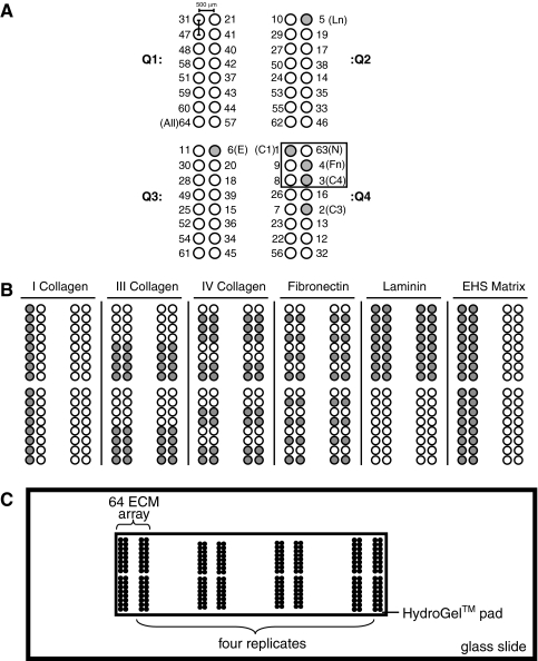
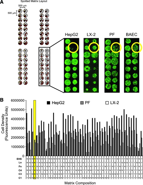
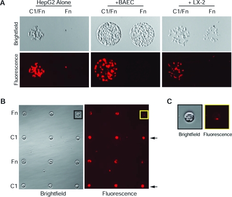
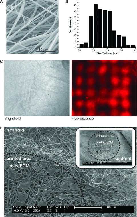
The spatial organization of cells of different phenotypes is an important and often defining determinant of tissue function. In tissue engineering, which attempts to rebuild functional tissues from cellular and synthetic components, spatial patterning of cells onto biomaterials is likely to be equally important. We have printed combinatorial arrays of extracellular matrix (ECM) and screened them for attachment by HepG2 hepatocytes, LX-2 hepatic stellate cells, primary portal fibroblasts, and bovine aortic endothelial cells-cells selected as representative phenotypes found in adult liver. Differential cell attachment to the underlying matrix proteins allowed us to establish two-dimensional co-cultures of HepG2 with these non-parenchymal cell types. These general approaches were then translated to tissue engineering scaffolds where deposition of ECM proteins onto electrospun polylactide meshes resulted in patterned HepG2 cultures. We observed that the spatial organization of fibronectin deposits influenced HepG2 attachment and the establishment of co-cultures on our arrays. These micropatterned co-culture systems should serve as valuable tools for studying the soluble and insoluble signals involved in liver development, function, and disease.
Figures
Similar articles
-
Functional 3-D cardiac co-culture model using bioactive chitosan nanofiber scaffolds.Biotechnol Bioeng. 2013 Feb;110(2):637-47. doi: 10.1002/bit.24727. Epub 2012 Oct 5. Biotechnol Bioeng. 2013. PMID: 22991229
-
Fabrication of transferable micropatterned-co-cultured cell sheets with microcontact printing.Biomaterials. 2009 Oct;30(29):5427-32. doi: 10.1016/j.biomaterials.2009.06.033. Epub 2009 Jul 16. Biomaterials. 2009. PMID: 19608271
-
Functional Maturation of Induced Pluripotent Stem Cell Hepatocytes in Extracellular Matrix-A Comparative Analysis of Bioartificial Liver Microenvironments.Stem Cells Transl Med. 2016 Sep;5(9):1257-67. doi: 10.5966/sctm.2015-0235. Epub 2016 Jul 15. Stem Cells Transl Med. 2016. PMID: 27421950 Free PMC article.
-
Galactose-carrying polymers as extracellular matrices for liver tissue engineering.Biomaterials. 2006 Feb;27(4):576-85. doi: 10.1016/j.biomaterials.2005.06.008. Epub 2005 Aug 8. Biomaterials. 2006. PMID: 16084586 Review.
-
Microporous membrane-based liver tissue engineering for the reconstruction of three-dimensional functional liver tissues in vitro.Biomatter. 2012 Oct-Dec;2(4):290-5. doi: 10.4161/biom.22481. Biomatter. 2012. PMID: 23507893 Free PMC article. Review.
Cited by
-
Therapeutic potential of stem cell in liver regeneration.Front Med. 2011 Mar;5(1):26-32. doi: 10.1007/s11684-011-0107-0. Epub 2011 Mar 17. Front Med. 2011. PMID: 21681671 Review.
-
Endothelial cell micropatterning: methods, effects, and applications.Ann Biomed Eng. 2011 Sep;39(9):2329-45. doi: 10.1007/s10439-011-0352-z. Epub 2011 Jul 15. Ann Biomed Eng. 2011. PMID: 21761242 Free PMC article. Review.
-
Bioengineered Liver Models for Drug Testing and Cell Differentiation Studies.Cell Mol Gastroenterol Hepatol. 2017 Dec 6;5(3):426-439.e1. doi: 10.1016/j.jcmgh.2017.11.012. eCollection 2018 Mar. Cell Mol Gastroenterol Hepatol. 2017. PMID: 29675458 Free PMC article. Review.
-
Genetic characteristics of the human hepatic stellate cell line LX-2.PLoS One. 2013 Oct 8;8(10):e75692. doi: 10.1371/journal.pone.0075692. eCollection 2013. PLoS One. 2013. PMID: 24116068 Free PMC article.
References
-
- Musat A.I. Sattler C.A. Sattler G.L. Pitot H.C. Reestablishment of cell polarity of rat hepatocytes in primary culture. Hepatology. 1993;18:198. - PubMed
-
- Moghe P.V. Coger R.N. Toner M. Yarmush M.L. Culture matrix configuration and composition in the maintenance of hepatocyte polarity and function. Biomaterials. 1996;17:373. - PubMed
-
- Berthiaume F. Moghe P.V. Toner M. Yarmush M.L. Effect of extracellular matrix topology on cell structure, function, and physiological responsiveness: hepatocytes cultured in a sandwich configuration. FASEB J. 1996;10:1471. - PubMed
-
- Bhandari R.N.B. Riccalton L.A. Lewis A.L. Fry J.R. Hammond A.H. Tendler S.J.B. Shakesheff K.M. Liver tissue engineering: a role for co-culture systems in modifying hepatocyte function and viability. Tissue Eng. 2001;7:345. - PubMed
-
- Bhatia S.N. Balis U.J. Yarmush M.L. Toner M. Microfabrication of hepatocyte/fibroblast co-cultures: role of homotypic cell interactions. Biotechnol Progr. 1998;14:378. - PubMed
Publication types
MeSH terms
Substances
Grants and funding
LinkOut - more resources
Full Text Sources
