. 2008 Dec;8(23-24):4919-30.
doi: 10.1002/pmic.200800608.

Two-dimensional gel-based approaches for the assessment of N-Linked and O-GlcNAc glycosylation in human and simian immunodeficiency viruses

Affiliations

Two-dimensional gel-based approaches for the assessment of N-Linked and O-GlcNAc glycosylation in human and simian immunodeficiency viruses

David R M Graham et al. Proteomics. 2008 Dec.

Abstract

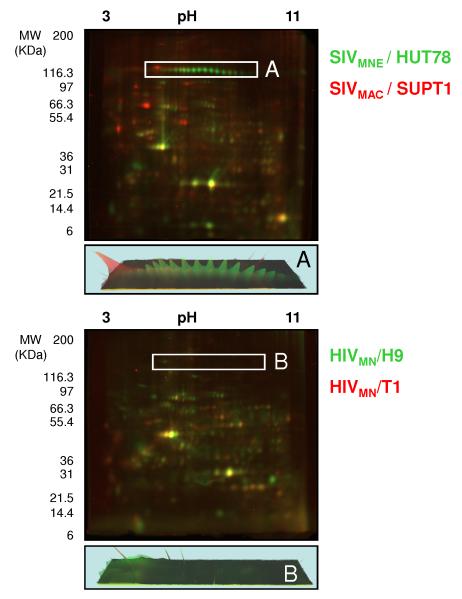
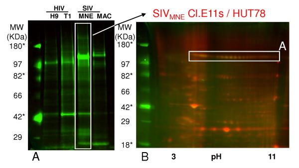
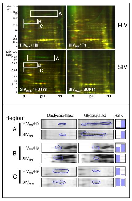
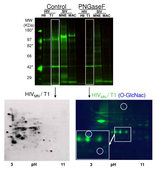
The glycosylation state of envelope glycoproteins in human and simian immunodeficiency viruses (HIV/SIV) is critical to viral infectivity and tropism, viral protein processing, and in virus evasion of the immune system. Using a rapid fluorescent 2-D gel-based method coupled with enzymatic pre-treatment of virus with PNGase F (Peptide: N-Glycosidase F) and fluorescent 2-D gels or 2-D gel Western blotting, we show significant differences in the glycosylation patterns of two SIV strains widely used in animal models of HIV disease and vaccine studies. We also demonstrate the modification of a host protein important in HIV biology (HLA-DR) by O-GlcNAc. Further, this experimental pipeline allows for the identification of the modified protein and the site of N-linked glycosylation by fluorescent 2-DE coupled with MS and the qualitative and semi-quantitative assessment of viral glycosylation. The method is fully compatible with downstream glycomics analysis. This approach will permit correlation of virus glycosylation status with pathological severity and may serve as a rapid screen of viruses from physiological samples for further study by more advanced MS methodology.


Figures

Figure 1
Figure 1. Comparison of SIV and HIV strains, illustrating the differences in viral and host protein composition
Two-dimensional gel electrophoresis with fluorescent labeling of 50μg of capsid protein equivalents for each virus was used to compare SIVMNE /HUT78, Cy3-labeled (green), and SIVMAC / SUPT1, Cy5-labeled (red) (top panel) and HIVMN / H9, Cy3-labeled (green), and HIVMN / T1, Cy5-labeled (red) (bottom panel). The region containing gp120 in SIV and HIV is indicated as box 1 and 2 respectively, and the representative three-dimensional protein abundance plot is presented for each region below. Gels were visualized by fluorescent scanning.
Figure 2
Figure 2. Pro-Q emerald staining (general carbohydrate stain) demonstrating the degree of glycosylation in HIV and SIV by 1 and 2-dimensional gel electrophoresis
Virus was normalized to capsid protein, and subjected to one-dimensional electrophoresis (left panel, approximately 10 μg of total protein each per lane), followed by staining with Pro-Q emerald to show differences in glycosylation. Notation is truncated. In the right panel, 15 μg of SIVMNE was labeled with Cy5, mixed with 35μg of unlabelled sample and subjected to two-dimensional gel electrophoresis followed by staining with Pro-Q emerald. Gels were visualized by fluorescent scanning. Region A corresponds to region A in Figure 1. * - represents molecular weight markers that are glycosylated.
Figure 3
Figure 3. PNGase F treatment of HIV and SIV and assessment by fluorescent 2-dimensional gel electrophoresis
Equal amounts of either HIV or SIV (strain indicated in white inlaid) were first treated with Peptide N-Glycosidase F (PNGase F) or with control buffers, labeled with either Cy3-control (green) or Cy5-PNGase F (red), and then subjected to two-dimensional electrophoresis (15 μg of each, for a total of 30μg per gel). Deglycosylation by PNGase F results in a decrease in molecular mass, and an acidic shift if the N-linked glycan is neutral (conversion of asparagines to aspartic acid). Gels were visualized by fluorescent scanning. In the bottom panel, regions identified as gp120 were analyzed by PROGENESIS software (see methods) and relative ratios of signal between glycosylated and deglycosylated gels were compared.
Figure 4
Figure 4. PNGase F removal of N-Linked Glycans and subsequent visualization of O-GlcNAc modified proteins in HIVMN/T1 by standard and fluorescent Western Blotting
The results from figure 2 are shown with the addition of results for PNGase F treatment of the same samples (PNGase F). Control virus (HIVMN/T1, 50 μg) was subjected to two-dimensional electrophoresis and transferred to PVDF. The blot was then probed with antibodies specific to O-GlcNAc and developed with an anti-mouse IgM-HRP conjugate (bottom left panel). Alternatively, following removal of N-linked glycans, HIVMN/T1 was Cy3-labeled (Green, 50 μg) and subjected to two-dimensional electrophoresis and then transferred to low-background PVDF. The blot was then probed with antibodies specific to OGlcNAc and subsequently with Cy2-labeled (blue) anti-mouse IgM. The blot was visualized by fluorescent scanning (bottom right panel).

References

    1. Chen M, Shi C, Kalia V, Tencza SB, et al. HIV gp120 V(1)/V(2) and C(2)-V(3) domains glycoprotein compatibility is required for viral replication. Virus Res. 2001;79:91–101. - PubMed
    1. Geijtenbeek TB, van Kooyk Y. DC-SIGN: a novel HIV receptor on DCs that mediates HIV-1 transmission. Curr Top Microbiol Immunol. 2003;276:31–54. - PubMed
    1. Land A, Braakman I. Folding of the human immunodeficiency virus type 1 envelope glycoprotein in the endoplasmic reticulum. Biochimie. 2001;83:783–790. - PubMed
    1. Kwong PD, Doyle ML, Casper DJ, Cicala C, et al. HIV-1 evades antibody-mediated neutralization through conformational masking of receptor-binding sites. Nature. 2002;420:678–682. - PubMed
    1. Shi Y, Brandin E, Vincic E, Jansson M, et al. Evolution of human immunodeficiency virus type 2 coreceptor usage, autologous neutralization, envelope sequence and glycosylation. J Gen Virol. 2005;86:3385–3396. - PubMed

Publication types

MeSH terms