. 2009 Mar;31(3):371-80.
doi: 10.1002/hed.20968.

Tumor-derived microvesicles in sera of patients with head and neck cancer and their role in tumor progression

Affiliations

Tumor-derived microvesicles in sera of patients with head and neck cancer and their role in tumor progression

Christoph Bergmann et al. Head Neck. 2009 Mar.

Abstract

Background: Tumor-derived membranous vesicles (MV) isolated from sera of the patients with squamous cell carcinomas of the head and neck (HNSCC) induce apoptosis of activated CD8(+) T cells. We tested if MV molecular profile and activity correlate with disease progression.

Methods: CD8(+) Jurkat cells were incubated with MAGE 3/6(+), FasL(+), MHC class I(+) MV isolated from sera of 60 patients with HNSCC and 25 normal controls by exclusion chromatography and ultracentrifugation. Z-VAD-FITC binding to Jurkat was measured and correlated with clinical data.

Results: MV from patients' sera, but not from sera of normal controls, induced Jurkat cell apoptosis. Forty-five percent T cells+MV from patients with N(1)-N(3) disease were apoptotic versus 18% T cells+MV from patients with N(0) disease (p < .008). MV from patients with active disease (AD) expressed higher FasL levels than MV from patients with no evident disease (NED) or normal controls (p <or= .01).

Conclusion: MAGE 3/6(+), FasL(+), and MHCI(+) MV in sera of patients induced T-cell apoptosis, which correlated with disease activity and the presence of lymph node metastases.


Figures
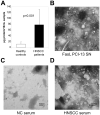
Figure 1
Figure 1. Microvesicles (MV) present in sera of HNSCC patients and normal controls
In A, protein content/10mL of serum in HNSCC patients and normal controls (NC). Microvesicles were isolated by exclusion chromatography and ultracentrifugation, as described in Materials and Methods. In B-D, transmission electron microscopy of negatively stained microvesicles fractionated from supernatants of FasL+ PCI-13 cells (B); from serum of a representative normal control (C); and from serum of a representative patient with HNSCC (D). The bar indicates 100nm.
Figure 2
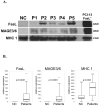
Figure 2. The molecular profile of microvesicles isolated from sera
In A, Western blot analysis of microvesicles for expression of FasL, MAGE 3/6 and MHC class I (MHC1) in fractions purified from sera of normal controls (NC), HNSCC patients (P1 – P5) and supernatants of a FasL+ PCI-13 cell line. Representative blots are shown. The numbers on the right indicate molecular weights of the detected proteins. In B, box-plots display distribution of results from semiquantitative immunoblots analysis showing a median as well as the first & third quartile, including outliers.
Figure 3
Figure 3. MHC class I and FasL expression levels in microvesicles isolated from sera of HNSCC patients with active disease, no evident disease and normal controls
In A, the percent of patients with serum-derived miscrovesicles which show low or high levels of MHC class 1 expression. Results are shown for patients with active disease (AD, n=48) or no evident disease (NED, n=11). In B, the percent of patients with serum-derived microvesicles which show high or low levels of the membrane form (42kDa) of FasL. In C, levels of FasL expression semiquantified based on densitometry readings and presented in relative units for microvesicles obtained from patients with active disease (AD), no evident disease (NED) or normal controls (NC). The p-values indicate a significant difference between AD and NED patients. Corresponding fractions of sera obtained from NC (in A or B) contained none or low levels of MHC I or FasL.
Figure 4
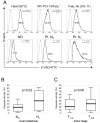
Figure 4. Pancaspase activation in CD8+ Jurkat cells co-incubated with microvesicles derived from sera of patients with HNSCC
In A, Z-VAD FMK binding to CD8+ Jurkat cells after treatment with heat or co-incubation with microvesicles of different origin, or with CH-11 antibody used as a positive control. Cells were co-incubated with microvesicles for 6h at 37°C in 5% CO2 in air, or treated for 30min at 56°C. Z-VAD-binding in Jurkat cells indicates pan-caspase activation which is taken as a sign of apoptosis. Upper row: in control experiments, caspase activation after the treatment with 56°C, FasL Ab, or microvesicles isolated from FasL+ PCI-13 SN. Lower row: representative blots show Z-VAD-binding in Jurkat cells after incubation with microvesicles from normal controls or a representative HNSCC patient without (N0) or with (N1) local metastases. In B, box plots summarize biologic activity of microvesicles derived from patients with (n=32) or without (n=28) lymph node metastase. MV biologic activity was determined measuring pan-caspase activation in CD8+ Jurkat cells after co-incubation with microvesicles for 6h. In C, box plots show biologic activity of microvesicles derived from patients with T1/2 (n=24) vs. T3/4 (n=36).

References

    1. Young MR. Protective mechanisms of head and neck squamous cell carcinomas from immune assault. Head Neck. 2006;28:462–470. - PubMed
    1. Grandis JR, Falkner DM, Melhem MF, Gooding WE, Drenning SD, Morel PA. Human leukocyte antigen class I allelic and haplotype loss in squamous cell carcinoma of the head and neck: clinical and immunogenetic consequences. Clin Cancer Res. 2000;6:2794–2802. - PubMed
    1. Whiteside TL. Immunobiology of head and neck cancer. Cancer Metastasis Rev. 2005;24:95–105. - PubMed
    1. Kim JW, Tsukishiro T, Johnson JT, Whiteside TL. Expression of pro- and antiapoptotic proteins in circulating CD8+ T cells of patients with squamous cell carcinoma of the head and neck. Clin Cancer Res. 2004;10:5101–5110. - PubMed
    1. Whiteside TL. Immune suppression in cancer: effects on immune cells, mechanisms and future therapeutic intervention. Semin Cancer Biol. 2006;16:3–15. - PubMed

Publication types

MeSH terms

Substances