. 2009 Mar 1;327(1):24-33.
doi: 10.1016/j.ydbio.2008.11.017. Epub 2008 Dec 3.

The human placenta is a hematopoietic organ during the embryonic and fetal periods of development

Affiliations

The human placenta is a hematopoietic organ during the embryonic and fetal periods of development

Alicia Bárcena et al. Dev Biol. .

Retraction in

Abstract

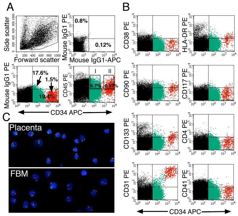
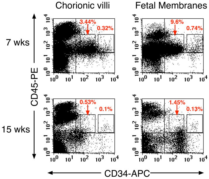
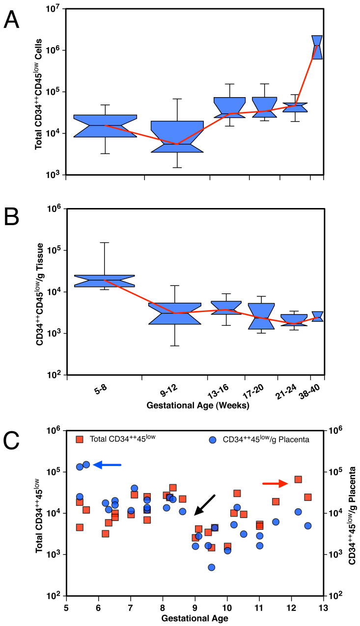
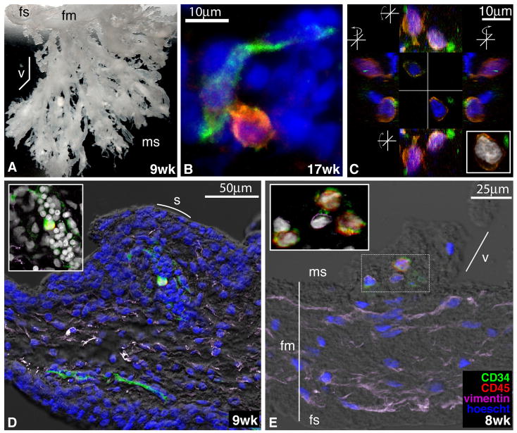
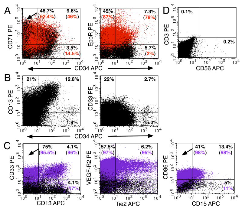
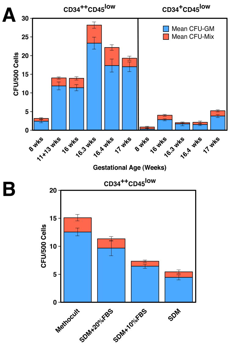
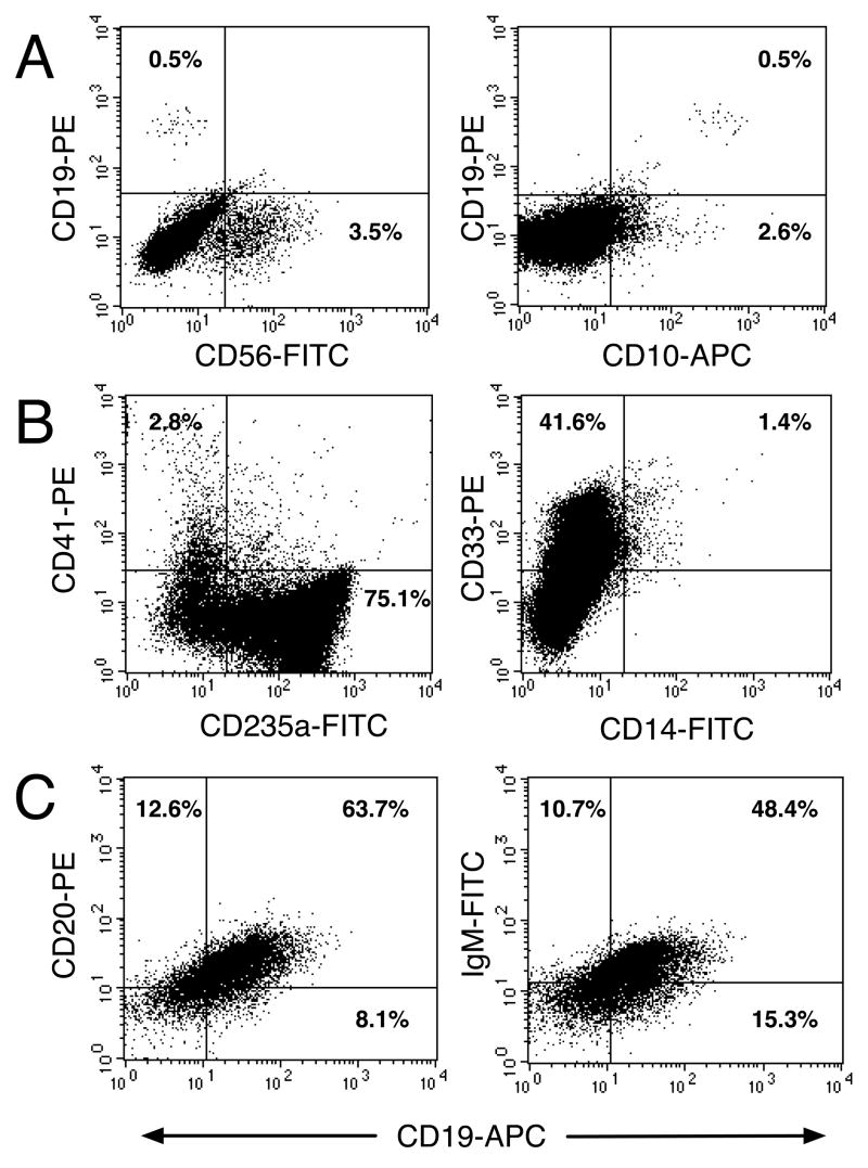
We studied the potential role of the human placenta as a hematopoietic organ during embryonic and fetal development. Placental samples contained two cell populations-CD34(++)CD45(low) and CD34(+)CD45(low)-that were found in chorionic villi and in the chorioamniotic membrane. CD34(++)CD45(low) cells express many cell surface antigens found on multipotent primitive hematopoietic progenitors and hematopoietic stem cells. CD34(++)CD45(low) cells contained colony-forming units culture (CFU-C) with myeloid and erythroid potential in clonogenic in vitro assays, and they generated CD56(+) natural killer cells and CD19(+)CD20(+)sIgM(+) B cells in polyclonal liquid cultures. CD34(+)CD45(low) cells mostly comprised erythroid- and myeloid-committed progenitors, while CD34(-) cells lacked CFU-C. The placenta-derived precursors were fetal in origin, as demonstrated by FISH using repeat-sequence chromosome-specific probes for X and Y. The number of CD34(++)CD45(low) cells increased with gestational age, but their density (cells per gram of tissue) peaked at 5-8 wk, decreasing more than sevenfold at the onset of the fetal phase (9 wk of gestation). In addition to multipotent progenitors, the placenta contained myeloid- and erythroid-committed progenitors indicative of active in situ hematopoiesis. These data suggest that the human placenta is an important hematopoietic organ, raising the possibility of banking placental hematopoietic stem cells along with cord blood for transplantation.


Conflict of interest statement

Conflict of interest: The authors have declared that no conflict of interest exists.

Figures

Figure 1
Figure 1
Phenotypic profile and fetal origin of placental hematopoietic progenitors. In A and B, freshly isolated light-density cell suspension from a 10-week placenta was stained with the indicated mAbs and PI, to identify dead cells. (A) The forward versus side-scatter profile of 105 cells and the electronic gate used to include all hematopoietic cells. Another gate was set to exclude PI+ cells (not shown). Isotype-matched negative controls are included. The gates set for cell sorting experiments used in C and in Figures 6 and 7 are shown in the CD34-APC versus CD45-PE dot plot (I for CD34+CD45low; II for CD34++CD45low). (B) Staining of the same cells with mAbs against markers expressed by hematopoietic progenitors. Multicolor analysis was performed to highlight the CD34++CD45low (red) and CD34+CD45low (green) cells. Results are representative of 12 experiments. (C) Sorted CD34++CD45low cells (using gate II in A) from placenta and from fetal BM (FBM) from the same 19-week specimen were hybridized with fluorescent probes specific for the X (green) or Y (red) chromosomes. Cell nuclei were stained with 4′,6′-diamidino-2-phenylindole (blue). Photomicrographs of the slides at 40x magnification were taken with a Nikon E-800 microscope/camera.
Figure 2
Figure 2
Hematopoietic progenitors were present in chorionic villi and the chorioamniotic membranes. Freshly isolated light-density cells from villi and fetal membranes (A, pooled 6.3 + 6.5 weeks; B, 15.3 weeks) were stained with CD34, CD45 and PI and analyzed (see Figure 1 legend).
Figure 3
Figure 3
The hematopoietic compartment of the placenta changes during gestation. (A) Box plot of total CD34++CD45 low placental cells grouped by gestational age (in weeks). (B) Box plot of the number of CD34++CD45low cells per gram of tissue grouped by gestational age. Box plots display the upper and lower quartiles surrounding the median (indicated by the notch), and the span of the whiskers shows the range of the data. Diamonds indicate the position of the mean values. Box width is proportional to sample size (5–8 weeks, n=18; 9–12 weeks, n=15; 13–16 weeks, n=9; 17–20 weeks, n=7; 21–24 weeks, n=7; 38–40 weeks, n=3). (C) Total numbers of CD34++CD45 low placental cells (red squares) and CD34++CD45 low placental cells per gram (blue circles) of individual 5-to 12-week tissues (n = 30). Blue arrow indicates the high number of CD34++CD45 low cells obtained per gram of 5-week tissues. Black arrow indicates the decrease at 9 weeks in total numbers and cells/gram tissue of CD34++CD45 low cells. Red arrow indicates the increase in total CD34++CD45 low placental cells at the end of the first trimester.
Figure 4
Figure 4
Localization and morphology of CD34+CD45+ cells within the human placenta. (A) Chorionic villi biopsied from a 9-week human placenta embedded in gelatin to demonstrate the macroscopic anatomy of these structures (2x magnification). fs, fetal side; fm, fetal membranes; v, villus; ms, maternal side. (B) CD34+CD45+ cell within the villous mesenchyme in close contact with a CD34+CD45 _endothelial cell. Tissue sections of the chorionic villi at the gestational ages indicated were stained with anti-CD34 (green) and anti-CD45 (red) mAbs, along with TOTO-3 iodide (blue). Confocal images were obtained by sequential scanning at 63x magnification. (C) Orthogonal views of two CD34+CD45+ cells, stained as in B and from the same 17-week placenta, demonstrating their close associations with CD34+CD45 cells. The Z projection (inset) shows the large size and complex morphology of the putative progenitors’ nuclei. (D) Longitudinal section of a villus showing a single CD34+CD45+ cell surrounded by a cluster of CD34+ CD45 cells. s, syncytium. The tissue section was stained with anti-CD34 (green), anti-CD45 (red), and anti-vimentin (purple) mAbs, Hoescht (blue), and visualized with differential interference contrast (DIC, gray scale). (E) Section of a young villus near the fetal membranes with CD34+CD45+ cells in the center (stained as in D). The Z projection (inset) demonstrates a cluster of CD34+CD45+ cells that appeared to be tethered to their vimentin-positive neighbors.
Figure 5
Figure 5
Erythroid and myeloid progenitors together with NK cells were present in the placenta throughout gestation. (A) A freshly isolated light-density cell suspension from a 21-week placenta was stained for erythroid markers and analyzed (see Figure 1 legend). Red indicates cells stained with an anti-CD235a-FITC (glycophorin A) mAb. (B) Analysis of a 12-week light-density placental cell suspension using mAbs that recognized myeloid progenitors in combination with an anti-CD34 mAb. (C) Four-color analysis of antigen expression on mature myeloid CD14+ cells (purple) (same cells as in B). (D) Analysis of CD3 and CD56 expression on 22-week placental cells. In all experiments, 1–2 × 10⁵ PI cells were analyzed. Results are representative of 5 experiments.
Figure 6
Figure 6
Colony-forming activity of placental hematopoietic progenitors. (A) Sorted populations from the placentas at the indicated gestational ages were plated in Methocult and scored at 3.5 weeks of culture. Results are shown as the mean ± SEM of 8–11 replicates for 4 experiments. CFU-GM, myeloid colonies. CFU-Mix, colonies containing both myeloid and erythroid cells. (B) Effects of FBS on colony-forming activity of CD34++CD45low sorted cells from a 19-week placenta. Cells were grown in commercial Methocult or in SDM supplemented with FBS.
Figure 7
Figure 7
CD34++CD45+ placental cells have multilineage hematopoietic potential. CD34++CD45low cells sorted from a 19-week placenta were cultured at 7.5 × 10³ cells/well in 48-well plates in SDM supplemented with SCF+FL+IL-7+IL-15+GM-CSF+IL-3 (A) or SCF+FL+Epo+GM-CSF+TPO (B). Viable cells (5 × 10⁴) were analyzed after 23 days in culture (see Figure 1 legend). Results are representative of 3 experiments. (C) CD34++CD45low cells sorted from a 12-week placenta were cultured at 5 × 10⁴ cells/well in 48-well plates with MS-5 murine BM stromal cells as feeders. After 3 weeks in culture, cells were detached, washed and stained with the indicated mAbs. The results of an analysis using a gate that contained viable CD45+FITC cells (left) and CD45+PE cells (right) are shown.

References

    1. Alvarez-Silva M, Belo-Diabangouaya P, Salaun J, Dieterlen-Lievre F. Mouse placenta is a major hematopoietic organ. Development. 2003;130:5437–5444. - PubMed
    1. Bárcena A, Muench MO, Galy AHM, Cupp J, Roncarolo MG, Phillips JH, Spits H. Phenotypic and functional analysis of T-cell precursors in the human fetal liver and thymus. CD7 expression in the early stages of T-and myeloid-cell development. Blood. 1993;82:3401–3414. - PubMed
    1. Bárcena A, Muench MO, Song KS, Ohkubo T, Harrison MR. Role of CD95/Fas and its ligand in the regulation of the growth of human CD34++CD38− fetal liver cells. Exp Hematol. 1999;27:1428–1439. - PubMed
    1. Bárcena A, Park SW, Banapour B, Muench MO, Mechetner EB. Expression of Fas/CD95 and bcl-2 in primitive hematopoietic progenitors in the human fetal liver. Blood. 1996;88:2013–2025. - PubMed
    1. Benischke K, Kaufmann P. Pathology of the Human Placenta. Springer; New York: 2000. Hofbauer cells; pp. 81–85.

Publication types