. 2009 Mar 30;178(1):75-9.
doi: 10.1016/j.jneumeth.2008.11.014. Epub 2008 Nov 27.

A technique for stereotaxic recordings of neuronal activity in awake, head-restrained mice

Affiliations

A technique for stereotaxic recordings of neuronal activity in awake, head-restrained mice

Jeri L Bryant et al. J Neurosci Methods. .

Abstract

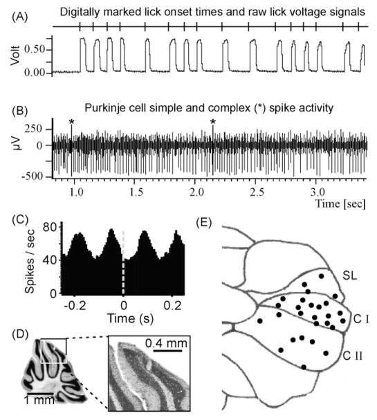
Neurophysiological recordings of brain activity during behavior in awake animals have traditionally been performed in primates because of their evolutionary close relationship to humans and comparable behavioral skills. However, with properly designed behavioral tasks, many fundamental questions about how the brain controls behavior can also be addressed in small rodents. Today, the rapid progress in mouse neurogenetics, including the development of mouse models of human brain disorders, provides unique and unparalleled opportunities for the investigation of normal and pathological brain function. The development of experimental procedures for the recording of neuronal activity in awake and behaving mice is an important and necessary step towards neurophysiological investigation of normal and pathological mouse brain function. Here we describe a method for stereotaxic recordings of neuronal activity from head-restrained mice during fluid licking. Fluid licking is a natural and spontaneous behavior in rodents, which mice readily perform under head-restrained conditions. Using a head-restrained preparation allows recordings of well-isolated single units at multiple sites during repeated experimental sessions. Thus, a large number of neurons can be tested for their relationship with behavior and detailed spatial maps of behavior related neuronal activity can be generated as exemplified here with recordings from lick-related Purkinje cells in the cerebellum.


Figures

Fig. 1
Fig. 1
Line drawing of the experimental setup for extracellular recordings from the cerebellum of an awake head-restrained mouse during an experiment involving licking behavior. The recording chamber and headpost are embedded in acrylic cement and are firmly attached to the mouse’s skull (see text and Fig. 2). The top of the headpost is inserted into the headpost clamp and held in place by a set screw. The mouse’s body is covered with a loose fitting plastic half-tube (5 cm diameter, 10 cm long) to limit body movements. The half-tube is held in place with adhesive tape. A water spout is placed within tongue-reach (4–6 mm) in front of the mouse’s mouth. A multiple-electrode microdrive with integrated pre-amplifiers is shown with the electrode guiding tubes inserted into the recording chamber.
Fig. 2
Fig. 2
Arrangement of headpost and recording chamber on the mouse’s skull and detailed photographs of the headpost and headpost clamp. All drawings are true to scale. (A) Side view of the skull with mountings. The headpost and recording chamber are embedded in acrylic cement, which is anchored to the skull with three screws, two of which are visible in this side view (black arrowheads). A stereotaxic micromanipulator was used to position the caudo-medial corner of the headpost on top of bregma. The vertical dashed line indicates the anterior–posterior coordinates of bregma. The recording chamber was fashioned from a drinking straw and the bottom was shaped with small scissors to match the curvature of the skull bone. (B) Top view of the skull showing the location of all three skull screws in relation to the headpost and recording chamber. This view illustrates that the headpost cannot be mounted more rostral and thus, that the distance between the recording chamber and the post cannot be further increased. The bend in the post increased the space between the recording chamber and the headpost clamp to allow unhindered access with recording equipment (see Fig. 1). The skull bone at the bottom of the recording chamber was removed to provide access to the underlying brain area (cerebellum shown here) through the intact dura. (C) Bottom view and (D) side view of the headpost clamp. The metal plate at the wide end of the fixture was used to attach the clamp to stationary metal post, which was mounted onto the surface of the experimental table. In (D) the headpost is maximally inserted into the opening as it would be during an experiment. The bend serves as a mechanical stop and determines how deep the post can be maximally inserted. This allows the reliable reproduction of z-axis coordinates across experiments. (E) Enlarged top view of the headpost clamp with headpost (post contour marked with dotted line) held in the lower right corner by the set screw. (F) Enlarged view of the headpost, which was cut out of a 3 mm thick aluminum sheet. The bend added three millimeters of distance between the headpost clamp and the recording chamber and also determined how deep the post could be inserted into the clamping fixture. (G) A true-to-scale drawing of the headpost clamp and set screw (dashed outlines) and a mouse’s skull with headpost and chamber. The 5–7 mm distance between the posterior edge of the clamping fixture and the recording chamber were sufficient for accessing the recording chamber with an electrode microdrive.
Fig. 3
Fig. 3
Representation of licking behavior in single unit Purkinje cell activity. (A) Recording of licking behavior: vertical lines in the top trace mark the ascending slopes of the lick junction potential, which correspond to the onsets of tongue-to-waterspout contacts. The trace below is the lick junction potential raw signal. Whenever the tongue touched the waterspout a positive junction potential lasting for the duration of the contact could be recorded. Junction potential onset times (ascending slopes) were detected with a threshold algorithm and used as temporal aligns for the lick-triggered correlation analysis of neuronal spike activity. (B) Single unit Purkinje cell simple and complex spike activity recorded simultaneously with the licking signals in (A). Asterisks mark complex spikes. We were able to obtain stable recordings from well-isolated single units for up to 20 min. (C) Cross-correlation analysis of simple spike activity with licking revealed a rhythmic phase-locked modulation of simple spike activity with rhythmic fluid licking. (D) Nissl stained parasagittal slice of a mouse cerebellum showing an electrolytic lesion at a recording site. The lesion was placed at stereotaxic coordinates relative to the headpost and thus relative to bregma. The framed part of the left image is shown enlarged on the right. (E) Schematic drawing of the right cerebellar hemisphere seen from the top. Black dots mark locations where single unit Purkinje cells with lick-locked rhythmically modulated simple spike activity were recorded. SL = simple lobule, C I, C II = Crus I and II.

References

    1. Bengtsson F, Jorntell H. Ketamine and xylazine depress sensory-evoked parallel fiber and climbing fiber responses. J Neurophysiol. 2007;98:1697–705. - PubMed
    1. Boughter JD, Baird JD, Bryant JL, St John SJ, Heck DH. C57BL/6J and DBA/2J Mice Vary in Lick rate and ingestive microstructure. genes Brain Behav. 2007;6:619–27. - PubMed
    1. Cheron G, Gall D, Servais L, Dan B, Maex R, Schiffmann SN. Inactivation of calcium-binding protein genes induces 160 Hz oscillations in the cerebellar cortex of alert mice. J Neurosci. 2004;24:434–41. - PMC - PubMed
    1. Davis S, Bliss TV, Dutrieux G, Laroche S, Errington ML. Induction and duration of long-term potentiation in the hippocampus of the freely moving mouse. J Neurosci Methods. 1997;75:75–80. - PubMed
    1. Errington ML, Bliss TV, Morris RJ, Laroche S, Davis S. Long-term potentiation in awake mutant mice. Nature. 1997;387:666–7. - PubMed

Publication types

LinkOut - more resources