. 2009 Mar;164(1):45-56.
doi: 10.1016/j.molbiopara.2008.11.006. Epub 2008 Nov 27.

Characterization of a protective Escherichia coli-expressed Plasmodium falciparum merozoite surface protein 3 indicates a non-linear, multi-domain structure

Affiliations

Characterization of a protective Escherichia coli-expressed Plasmodium falciparum merozoite surface protein 3 indicates a non-linear, multi-domain structure

Chiawei W Tsai et al. Mol Biochem Parasitol. 2009 Mar.

Abstract

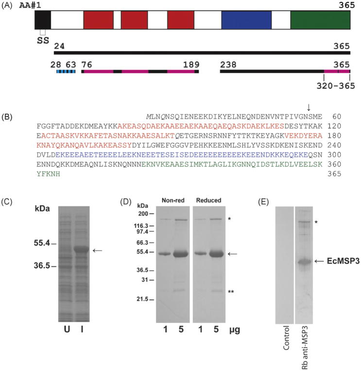
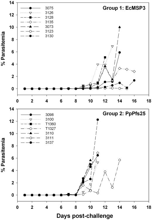
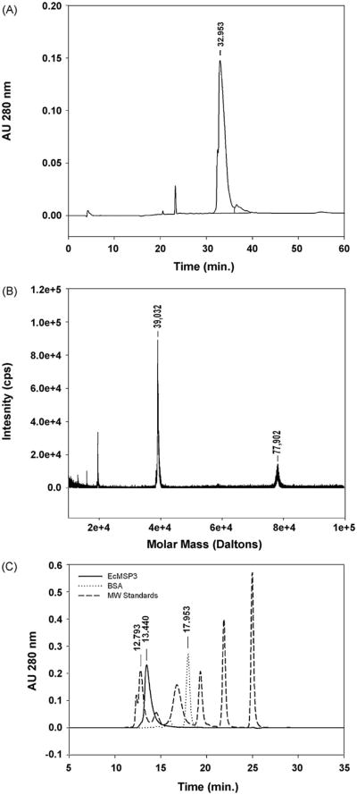
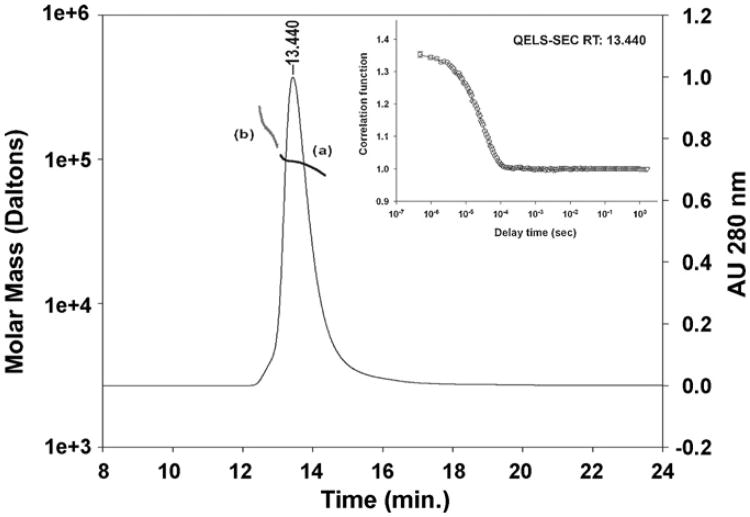
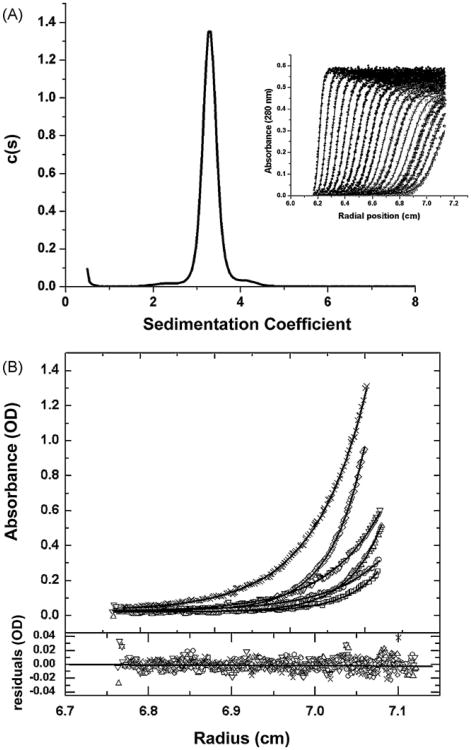
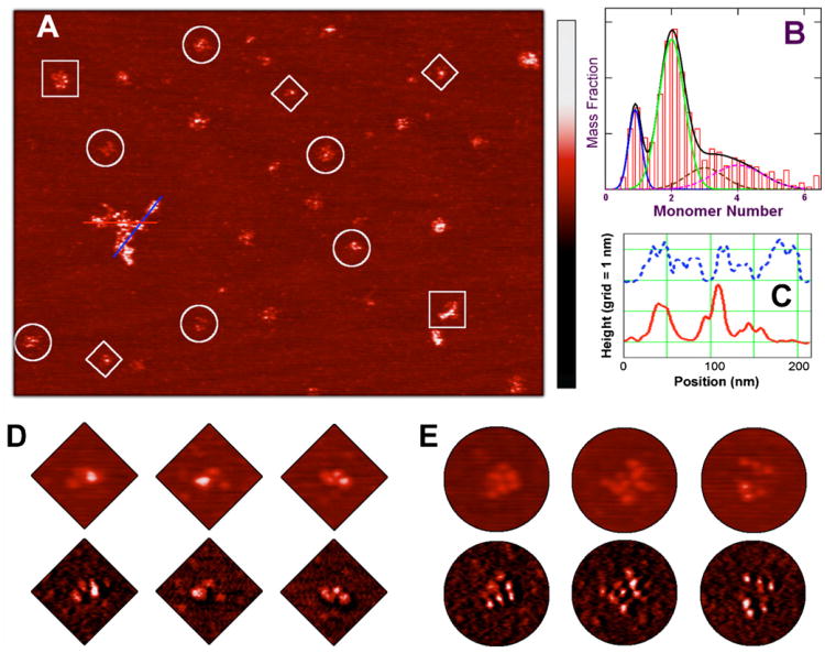
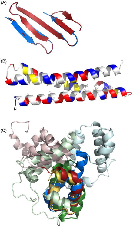
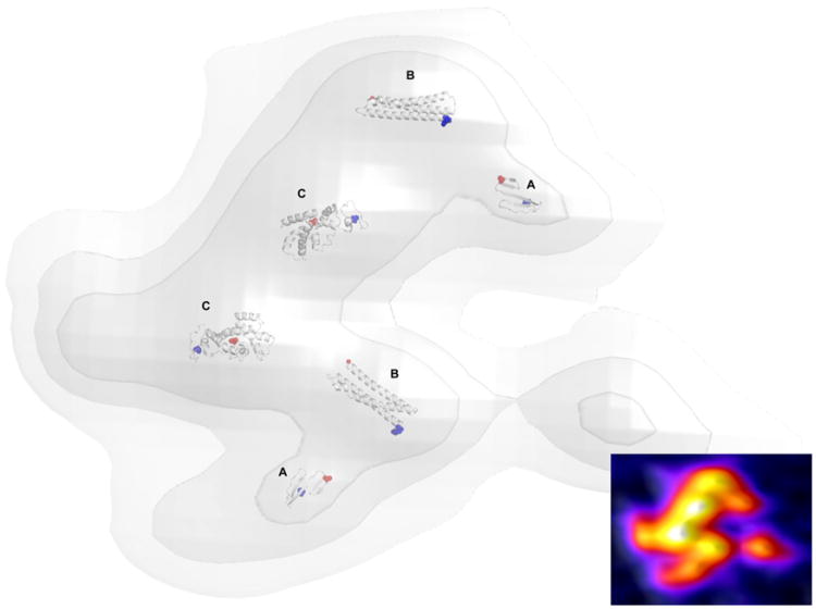
Immunization with a recombinant yeast-expressed Plasmodium falciparum merozoite surface protein 3 (MSP3) protected Aotus nancymai monkeys against a virulent challenge infection. Unfortunately, the production process for this yeast-expressed material was not optimal for human trials. In an effort to produce a recombinant MSP3 protein in a scaleable manner, we expressed and purified near-full-length MSP3 in Escherichia coli (EcMSP3). Purified EcMSP3 formed non-globular dimers as determined by analytical size-exclusion HPLC with in-line multi-angle light scatter and quasi-elastic light scatter detection and velocity sedimentation (R(h) 7.6+/-0.2nm and 6.9nm, respectively). Evaluation by high-resolution atomic force microscopy revealed non-linear asymmetric structures, with beaded domains and flexible loops that were recognized predominantly as dimers, although monomers and larger multimers were observed. The beaded substructure corresponds to predicted structural domains, which explains the velocity sedimentation results and improves the conceptual model of the protein. Vaccination with EcMSP3 in Freund's adjuvant-induced antibodies that recognized native MSP3 in parasitized erythrocytes by an immunofluorescence assay and gave delayed time to treatment in a group of Aotus monkeys in a virulent challenge infection with the FVO strain of P. falciparum. Three of the seven monkeys vaccinated with EcMSP3 had low peak parasitemias. EcMSP3, which likely mimics the native MSP3 structure located on the merozoite surface, is a viable candidate for inclusion in a multi-component malaria vaccine.


Figures

Fig. 1
Fig. 1
Production of EcMSP3. A schematic of the MSP3 protein (solid black line represents expressed region of MSP3, broken black line represents portions of the sequence used for structure modeling, cyan indicates beta strands and magenta indicates alpha helices) (A), amino acid sequence of EcMSP3 including non-native M (B), Coomassie blue stained SDS-PAGE gels with uninduced (U) and induced (I) solubilized cells produced by shake flask fermentation (C) and purified EcMSP3 protein (D), and analysis by immunoblot with rabbit anti-EcMSP3 antiserum or control (E). Blocks within the schematic as well as amino acids highlighted by colors are the three blocks (H1–H3) of Ala-X-X-Ala-X-X-X heptad repeats (red), the glutamic acid-rich region (blue), and the leucine zipper-like domain (green). Two glutamines (Q) shown in italics in panel B are N to Q point mutations introduced into the synthetic gene (AF440682).
Fig. 2
Fig. 2
Aotus challenge study. Parasitemia curves in Aotus monkeys challenged intravenously with P. falciparum-infected Aotus erythrocytes after immunization with EcMSP3 or PpPfs25, a control protein.
Fig. 3
Fig. 3
Biochemical analysis of EcMSP3. EcMSP3 was characterized by RP-HPLC (A), matrix assisted laser desorption ionization mass spectrometry (B) and SEC-HPLC (solid line) in comparison with molecular weight standards (dashed line) and bovine serum albumin (dash-dot-dash line) (C). Molecular weight standards used: thyroglobulin (670 kDa), immunoglobulin (158 kDa), ovalbumin (44 kDa), myoglobin (17 kDa) and vitamin B12 (1350 Da).
Fig. 4
Fig. 4
Biophysical characterization by inline SEC-MALS and QELS. Analysis of EcMSP3 by in-line MALS-SEC yielded the molar mass distribution of main peak (a, dark line, RT 13.440 min) and front shoulder (b, gray line) compared to the absorbance 280 nm. The insert shows SEC-QELS-HPLC result for the goodness of fit of the hydrodynamic radius at the apex of the primary peak (RT 13.440).
Fig. 5
Fig. 5
Biophysical characterization by sedimentation analyses. Panel A: sedimentation velocity characterization of EcMSP3. The inset of this figure shows the sedimentation boundary movement (time independent noise removed) of the EcMSP3 sample vs. radial position. The solid lines through the data points are the fit generated by Sedfit software analysis. For clarity only one half of the optical scans are shown, but data analysis was performed for all the scans. The deconvolution of the boundary data into resolved sedimenting components is shown as a c(s) vs. s plot (see Section 2). The essentially single peak was integrated to give a weight average sedimentation coefficient of 3.29S. Panel B: sedimentation equilibrium determination of the molar mass of MSP3. The top panel shows the overlay of the sedimentation equilibrium distributions, obtained at 4°C, absorbance vs. radius scans at 280 nm for EcMSP3 at the following concentrations: 0.22, 0.44, and 1.09 mg/mL at centrifugal speeds of 12,000 and 15,000 rpm. The × and diamond symbols show the data for the 1.09 mg/mL sample at 12,000 and 15,000 rpm, respectively. The inverted and upright triangles show the data for the 0.44 mg/mL sample at 12,000 and 15,000 rpm, respectively. The open circles and open squares show the data for the 0.22 mg/mL sample at 12,000 and 15,000 rpm, respectively. The solid line for each data set is the best fit to the data from the global analysis of all the scans using a single species model (see text). This non-linear regression analysis gives a molar mass 100,199 for EcMSP3. Residuals of the fitted lines to the experimental data are displayed in the lower panel with the corresponding symbols listed above. The best-fit rms error is 0.0058 and the global reduced χ2 was 2.0169.
Fig. 6
Fig. 6
Atomic force microscopy structures of EcMSP3 and oligomers. EcMSP3 and oligomers are highly resolved, after adsorption initiated under 20 mM PBS buffer (pH 7.4) over a flat mica surface pretreated with a CaCl2 solution, revealing large filamentous structures and smaller, isolated particles. Both structures have a consistent topological height of around 1.5 nm, but their sizes range from tens of nanometers to several hundred nanometers (A). The isolated particles can be grouped according to their volumes and identified as monomers, dimers, and higher order oligomers. The mass distribution derived from over 2000 EcMSP3 particles of high-resolution AFM images reveals discrete populations of monomer, dimer, and larger oligomers (B). The mass ratio of the dominant dimer population (under the green curve) to the monomer population (blue) is approximately four. The distribution of larger oligomers, totaling about 30% in the overall mass, can be attributed mainly as trimers (brown), tetramers (magenta) and a few sporadic higher oligomers. The solid black curve is the sum of the four components from monomer to tetramers. Each feature in the topological trace displays a beaded substructure as plotted on the line chart (C), showing the topological height along two cross sections of the largest structure in (A). The bead separation averaged about 14 nm. The number of resolved beads in the isolated particles corresponded with their oligomeric state. EcMSP3 monomers (diamonds) typically had three beads, dimers (circles) had up to seven to nine beads, and higher order oligomers (squares) had up to 12–15 beads. The color scale bar from dark to red to bright is universal with a range of 0–4 nm for all height images and of 0–15 degrees for all phase images.
Fig.7
Fig.7
Computational prediction of MSP3 structure. Structural models of MSP3 were generated by homology from sequence threading (LOOPP) or de novo with Rosetta. (A) Rosetta predicts that the N-terminal domain forms a small anti-parallel beta-sheet; two alternative models are shown in blue and red. (B) The alanine repeat region forms a coiled coil of three helices. These alanines face the interior of the bundle. Charged residues are shown in blue and red, hydrophobic residues in yellow (C) Rosetta predicts a leucine zipper-like domain at the C-terminus (shown in darker colors). This leucine zipper-like domain is a structural anchor around which four helices of the D/E-rich middle domain pack (shown in lighter colors). The top-scoring decoys from nearly 200,000 trials using the EcMSP3 sequence have little internal agreement and the top-scoring decoys from another 100,000 trials with closely related homologues do not agree between themselves or with the EcMSP3 decoys, suggesting that the orientation of these helices cannot be assigned with any confidence. The leucine zipper-like domain alone is shown in gold, the EcMSP3 C-terminus in red, the MSP3-K1 C-terminus in green, and the MSP3-NF54 C-terminus in blue.
Fig. 8
Fig. 8
A structural model of dimeric MSP3. The N-terminus and coiled coil regions are predicted with high confidence (A and B, respectively). The larger C-terminus contains leucine zipper-like domain responsible for dimerization (C). The top-scoring predictions for EcMSP3 were chosen for fitting to the protein pseudodensity map derived from high-resolution AFM topological imaging. The N-termini of each domain are colored blue and the C-termini of each domain are colored red. A heat map of the corresponding AFM image data is inset.

References

    1. Snow RW, Guerra CA, Noor AM, Myint HY, Hay SI. The global distribution of clinical episodes of Plasmodium falciparum malaria. Nature. 2005;434(7030):214–7. - PMC - PubMed
    1. McColl DJ, Silva A, Foley M, et al. Molecular variation in a novel polymorphic antigen associated with Plasmodium falciparum merozoites. Mol Biochem Parasitol. 1994;68(1):53–67. - PubMed
    1. Oeuvray C, Bouharoun-Tayoun H, Gras-Masse H, et al. Merozoite surface protein-3: a malaria protein inducing antibodies that promote Plasmodium falciparum killing by cooperation with blood monocytes. Blood. 1994;84(5):1594–602. - PubMed
    1. Singh S, Soe S, Mejia JP, et al. Identification of a conserved region of Plasmodium falciparum MSP3 targeted by biologically active antibodies to improve vaccine design. J Infect Dis. 2004;190(5):1010–8. - PubMed
    1. Roussilhon C, Oeuvray C, Muller-Graf C, et al. Long-term clinical protection from falciparum malaria is strongly associated with IgG3 antibodies to merozoite surface protein 3. PLoS Med. 2007;4(11):e320. - PMC - PubMed

Publication types

MeSH terms

LinkOut - more resources