. 2009 Feb 6;284(6):3619-27.
doi: 10.1074/jbc.M809269200. Epub 2008 Dec 10.

Interactions of HIV-1 inhibitory peptide T20 with the gp41 N-HR coiled coil

Affiliations

Interactions of HIV-1 inhibitory peptide T20 with the gp41 N-HR coiled coil

Kelly Champagne et al. J Biol Chem. .

Abstract

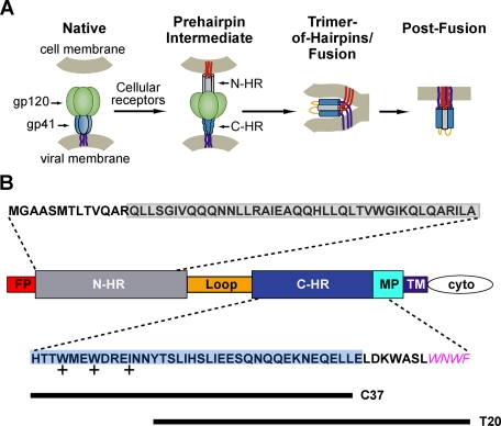
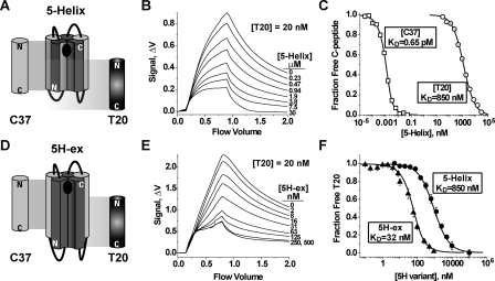
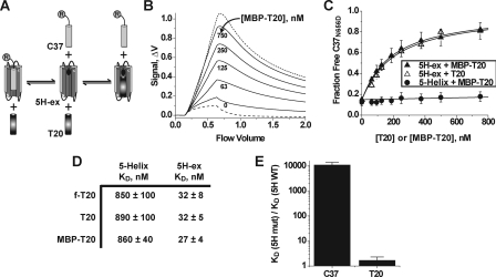
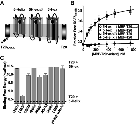
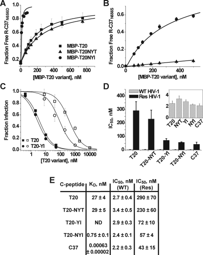
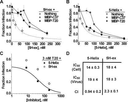
Cellular entry of human immunodeficiency virus type 1 (HIV-1) involves fusion of viral and cellular membranes and is mediated by structural transitions in viral glycoprotein gp41. The antiviral C-peptide T20 targets the gp41 N-terminal heptad repeat region (N-HR), blocking gp41 conformational changes essential for the entry process. To probe the T20 structure-activity relationship, we engineered a molecular mimic of the entire gp41 N-HR coiled coil using the 5-Helix design strategy. T20 bound this artificial protein (denoted 5H-ex) with nanomolar affinity (K(D) = 30 nm), close to its IC50 concentration (approximately 3 nm) but much weaker than the affinity of a related inhibitory C-peptide C37 (K(D) = 0.0007 nm). T20/C37 competitive binding assays confirmed that T20 interacts with the hydrophobic groove on the surface of the N-HR coiled coil outside of a deep pocket region crucial for C37 binding. We used 5H-ex to investigate how the T20 N and C termini contributed to the inhibitor binding activity. Mutating three aromatic residues at the T20 C terminus (WNWF --> ANAA) had no effect on affinity, suggesting that these amino acids do not participate in T20 binding to the gp41 N-HR. The results support recent evidence pointing to a different role for these residues in T20 inhibition (Peisajovich, S. G., Gallo, S. A., Blumenthal, R., and Shai, Y. (2003) J. Biol. Chem. 278, 21012-21017; Liu, S., Jing, W., Cheung, B., Lu, H., Sun, J., Yan, X., Niu, J., Farmar, J., Wu, S., and Jiang, S. (2007) J. Biol. Chem. 282, 9612-9620). By contrast, mutations near the T20 N terminus substantially influenced inhibitor binding strength. When Ile was substituted for Thr in the second T20 position, a 40-fold increase in binding affinity was measured (K(D) = 0.75 nm). The effect of this affinity enhancement on T20 inhibitory potency varied among different viral strains. The original T20 and the higher affinity T20 variant had similar potency against wild type HIV-1. However, the higher affinity T20 variant was significantly more potent against T20-resistant virus. The findings suggest that other factors in addition to binding affinity play a role in limiting T20 potency. As a mimetic of the complete gp41 N-HR coiled coil region, 5H-ex will be a useful tool to further elucidate mechanistic profiles of C-peptide inhibitors.


Figures

FIGURE 1.
FIGURE 1.
HIV-1 gp41 and its role in viral membrane fusion. A, a model of HIV-1 entry (46). In native Env prior to receptor activation, gp41 is held in a metastable conformation by a canopy of gp120 proteins (green). Receptor binding to gp120 stimulates gp41 to extend and insert its fusion peptide segment (red) into the target cell membrane. The N-HR (gray) and C-HR (blue) regions of the gp41 ectodomain are transiently exposed in this prehairpin state. Subsequently, gp41 collapses into the trimer-of-hairpins conformation that brings the gp41 fusion peptides, transmembrane regions (purple), and their associated membranes into the close proximity for membrane fusion. The actual disposition of gp120 in both the prehairpin and trimer-of-hairpins states is uncertain; for clarity, the protein is omitted in the schematic of the trimer-of-hairpins conformation. B, a diagram of HIV-1 gp41 identifying its fusion peptide (FP), N-HR, C-HR, MPER (MP), transmembrane (TM), and cytoplasmic (cyto) domains. Amino acid sequences above and below the diagram are derived from the N-HR and C-HR/MPER regions of EnvHXB2; all but the MPER sequence WNWF (magenta) were used in the design of 5H-ex. The N-HR and C-HR segments found in the original 5-Helix are boxed in gray and blue, respectively, whereas the sequences of C37 and T20 are denoted by lines. The side chains of the C-HR amino acids marked with + pack into the hydrophobic pocket at the C terminus of the N-HR coiled coil.
FIGURE 2.
FIGURE 2.
C-peptide binding properties of 5-Helix and 5H-ex. A and D, schematic diagrams of 5-Helix (A) and 5H-ex (D). The N-HR (dark gray) and C-HR (light gray) segments are shown as cylinders, whereas the Gly/Ser linkers are represented by black lines. The black oval in the C-peptide binding site indicates the location of the deep hydrophobic pocket at the N-HR C terminus. The cylinders labeled C37 and T20 show the extent of the binding site of each C-peptide. The N and C designate the N and C termini for each polypeptide. B and E, fluorescence response as equilibrated mixtures of f-T20 (20 nm), and either 5-Helix (B) or 5H-ex (E) were washed through the Kinexa 3000 flow fluorimeter. The instrument flow cell contained beads that specifically captured free (unbound) f-T20. The difference in fluorescence signals (Δf) measured before sample load (0 ml) and after the sample washout (2 ml) was proportional to the free f-T20 concentration in reaction mixtures (see “Experimental Procedures”). C and F, titrations of fluorescent C37 (2.5 pm) and T20 (20 pm) with either 5-Helix or 5H-ex. In C, R-C37 (squares) and f-T20 (circles) were titrated by 5-Helix, whereas in F, f-T20 was titrated by either 5-Helix (circles) or 5H-ex (triangles). The fraction of unbound C-peptide was calculated by normalizing each Δf to a minimum and maximum Δf obtained from samples in which the C-peptide is fully bound or fully unbound. The KD values were determined by fitting these data to a bimolecular equilibrium binding model (solid lines).
FIGURE 3.
FIGURE 3.
Properties of the 5H-ex/T20 interaction. A, overview of the C37 displacement assay. T20 and R-C37 compete for the same binding site on 5H-ex, and therefore titration of T20 (or MBP-T20) leads to an increase in the free (unbound) concentration of R-C37. The circled R denotes the rhodamine label linked to C37. B, Kinexa 3000 fluorescence response as solutions of 5H-ex (10 nm), R-C37N656D (1 nm), and MBP-T20 (indicated concentration) were passed over beads that specifically captured unbound R-C37N656D. The dashed and dotted lines were generated by control samples in which R-C37N656D is fully bound or fully unbound, respectively (see “Experimental Procedures”). The fraction of unbound R-C37N656D in each sample was calculated by normalizing the difference fluorescence signal Δf to the minimum and maximum Δf obtained from the control solutions. C, C37N656D displacement from 5H-ex (triangles) or 5-Helix (circles) as T20 (open symbols) or MBP-T20 (filled symbols) was titrated into reaction mixtures. The data points represent the means ± S.E. of at least three separate measurements and have been fit to a three-state competitive binding model (solid lines). D, tabulated KD values for the interaction of T20 with either 5-Helix or 5H-ex as determined using the direct binding assay (f-T20) or the C37 displacement assay (T20, MBP-T20). The values represent the means and S.E. of three or more separate measurements. E, effect of the W571R/G572D substitution in the N-HR hydrophobic pocket on C-peptide binding affinities. The data are represented as fold change in KD value relative to the wild type 5H-ex/C-peptide KD. The KD values were determined using the direct binding assay for C37 values or the C37 displacement assays for MBP-T20 values.
FIGURE 4.
FIGURE 4.
Role of the C-terminal C-HR/MPER sequence in stabilizing T20 binding to 5H-ex. A, schematic representations of 5-Helix, 5H-ex, and the slightly truncated 5H-exΔ5. The extent of the C-peptide binding site of each protein is indicated relative to T20 and T20ANAA peptides. B, C37N656D displacement measured using 5H-ex and MBP-T20ANAA (open circles), 5H-ex and MBP-T20 (filled circles), 5H-exΔ5 and MBP-T20 (open squares), and 5-Helix and MBP-T20 (filled diamonds). The data represent the means and S.E. of three or more independent experiments and were analyzed as described in the legend to Fig. 3. C, effect of C-peptide point mutations on the stability of the T20/5H-ex complex. Binding free energies were calculated using the formula ΔGo = -RTln(KD), where the KD values were obtained using the C37N656D displacement assay with MBP-T20 constructs as titrants. The horizontal lines indicate the binding energies of wild type MBP-T20 with either 5H-ex (solid) or 5-Helix (dotted).
FIGURE 5.
FIGURE 5.
Effect of the T639I substitution on T20 binding and inhibitory activities. A, C37N656D displacement from 5H-ex by MBP-T20 (squares), MBP-T20NYT (triangles), and MBP-T20NYI (circles). The data were obtained and analyzed as described in the legend to Fig. 3. B, as in A, except that the higher affinity C37N656S was used in place of C37N656D. Note that only the MBP-T20NYT (triangles) and MBP-T20NYI (circles) titrations are shown. C, titration of viral infectivity by T20 (squares) and T20YI containing the T639I substitution (circles). HIV-1 was pseudotyped with either wild type EnvHXB2 (filled symbols) or its mutant variant containing T20 escape mutations G547D/I548T (open symbols). The data represent the means ± S.E. of three to seven separate experiments and have been fit to a Langmuir function (solid lines) to determine IC50 (see “Experimental Procedures”). D, C-peptide IC50 values determined for the inhibition of HIV-1 psuedotyped with wild type Env (gray) or the G547D/I548T mutant variant (black). Inset, IC50 values for inhibition of wild type HIV-1 are plotted on an expanded scale. E, table of C-peptide IC50 and KD values. Res refers to the T20-resistant Env containing the G547D/I548T substitutions. ND, not determined.
FIGURE 6.
FIGURE 6.
Inhibitory properties of 5H-ex and 5-Helix. A and B, Titration of HIV-1 infectivity by 5H-ex (A) or 5-Helix (B) in the absence (open circles) or presence of 100 nm MBP-T20 (filled circles) or 100 nm MBP-C37 (squares). The data represent the mean ± S.E. from three or more separate experiments. The dotted line in both panels is a fit of the 5H-ex data to the Langmuir equation (see “Experimental Procedures”). C, titration of HIV-1 infectivity by 5H-ex (circles) or 5-Helix (squares) in the presence of 3 nm T20. The solid line depicts the expected infection level if the mixed inhibitors bound gp41 in a mutually exclusive manner and their antiviral activities were additive: Fraction infection = (1 + ([T20]/IC50, T20) + ([5H]/IC50, 5H))-1. D, tabulated inhibitory parameters for 5H-ex and 5-Helix. IC50 values are reported for cell-cell fusion (Cell) and HIV-1 infectivity (Virus) assays and represent the means and S.E. of three or more independent experiments. The CI determined for multiple mixtures of T20 and 5-Helix or 5H-ex was calculated as described under “Experimental Procedures.”

Similar articles

Cited by

References

    1. Peisajovich, S. G., Gallo, S. A., Blumenthal, R., and Shai, Y. (2003) J. Biol. Chem. 278 21012-21017 - PubMed
    1. Liu, S., Jing, W., Cheung, B., Lu, H., Sun, J., Yan, X., Niu, J., Farmar, J., Wu, S., and Jiang, S. (2007) J. Biol. Chem. 282 9612-9620 - PubMed
    1. Fenouillet, E., and Jones, I. M. (1995) J. Gen. Virol. 76 1509-1514 - PubMed
    1. Chan, D. C., Chutkowski, C. T., and Kim, P. S. (1998) Proc. Natl. Acad. Sci. U. S. A. 95 15613-15617 - PMC - PubMed
    1. Root, M. J., and Hamer, D. H. (2003) Proc. Natl. Acad. Sci. U. S. A. 100 5016-5021 - PMC - PubMed

Publication types

MeSH terms