. 2009 Feb 6;284(6):3537-45.
doi: 10.1074/jbc.M807001200. Epub 2008 Dec 9.

Oxidative stress alters syndecan-1 distribution in lungs with pulmonary fibrosis

Affiliations

Oxidative stress alters syndecan-1 distribution in lungs with pulmonary fibrosis

Corrine R Kliment et al. J Biol Chem. .

Abstract

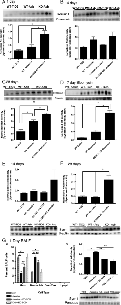
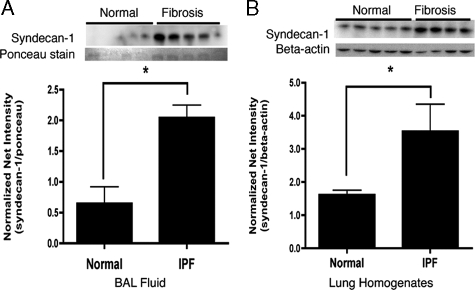
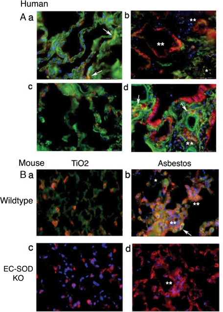
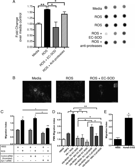
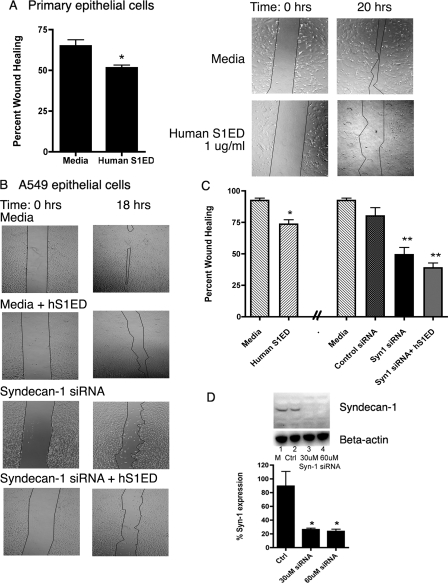
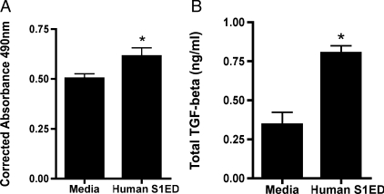
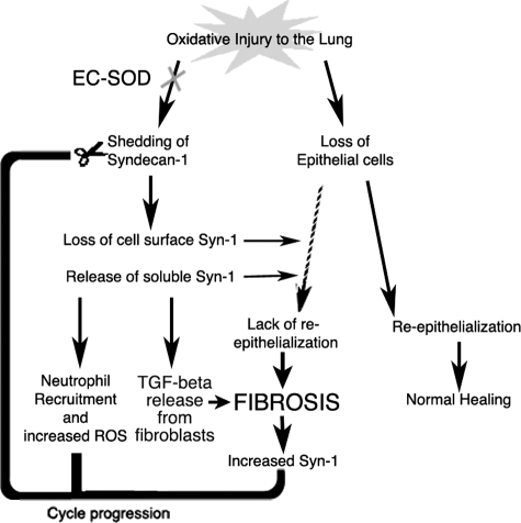
Idiopathic pulmonary fibrosis (IPF) is an interstitial lung disease characterized by severe, progressive fibrosis. Roles for inflammation and oxidative stress have recently been demonstrated, but despite advances in understanding the pathogenesis, there are still no effective therapies for IPF. This study investigates how extracellular superoxide dismutase (EC-SOD), a syndecan-binding antioxidant enzyme, inhibits inflammation and lung fibrosis. We hypothesize that EC-SOD protects the lung from oxidant damage by preventing syndecan fragmentation/shedding. Wild-type or EC-SOD-null mice were exposed to an intratracheal instillation of asbestos or bleomycin. Western blot was used to detect syndecans in the bronchoalveolar lavage fluid and lung. Human lung samples (normal and IPF) were also analyzed. Immunohistochemistry for syndecan-1 and EC-SOD was performed on human and mouse lungs. In vitro, alveolar epithelial cells were exposed to oxidative stress and EC-SOD. Cell supernatants were analyzed for shed syndecan-1 by Western blot. Syndecan-1 ectodomain was assessed in wound healing and neutrophil chemotaxis. Increases in human syndecan-1 are detected in lung homogenates and lavage fluid of IPF lungs. Syndecan-1 is also significantly elevated in the lavage fluid of EC-SOD-null mice after asbestos and bleomycin exposure. On IHC, syndecan-1 staining increases within fibrotic areas of human and mouse lungs. In vitro, EC-SOD inhibits oxidant-induced loss of syndecan-1 from A549 cells. Shed and exogenous syndecan-1 ectodomain induce neutrophil chemotaxis, inhibit alveolar epithelial wound healing, and promote fibrogenesis. Oxidative shedding of syndecan-1 is an underlying cause of neutrophil chemotaxis and aberrant wound healing that may contribute to pulmonary fibrosis.


Figures

FIGURE 1.
FIGURE 1.
Lack of EC-SOD leads to significant increases of syndecan-1 in bronchoalveolar lavage fluid (BALF) and lung tissue after asbestos or bleomycin exposure. Syndecan-1 in the BALF and lung homogenates of wild type and EC-SOD KO mice was detected by Western blot analysis and presented as normalized net intensity (mean ±S.E.), n = 5 mice per group. Results for the BALF data are standardized to protein loading as determined by Ponceau red staining of the membrane. Results from lung homogenates are standardized to β-actin. Shed syndecan-1 in the BALF at: (A) 1-day post-asbestos exposure, *, p < 0.05; (B) 14-days post-asbestos exposure, *, p < 0.05; (C) 28-days post-asbestos exposure, *, p < 0.05, **, p < 0.001; and (D) 7-days post-bleomycin exposure, *, p < 0.001. E, syndecan-1 in lung homogenates at 14-days post-asbestos exposure and (F) 28 days post-asbestos exposure, *, p < 0.05. Co-treatment of EC-SOD KO mice with asbestos and purified EC-SOD results in G–a decreased neutrophils, *, p < 0.001; **, p < 0.05; and in G-b decreased levels of syndecan-1, *, p < 0.05; **, p < 0.01, in the BALF at day 1, n = 4.
FIGURE 2.
FIGURE 2.
Syndecan-1 increases in the BALF and lung homogenates of IPF lungs compared with normal. Syndecan-1 was detected by Western blot analysis and presented as normalized net intensity (standardized to protein loading as determined by Ponceau red staining of the membrane for BALF samples and standardized to β-actin for lung homogenate samples (mean ±S.E.). Increased syndecan-1 in (A) BALF samples; *, p < 0.01, n = 5 and (B) lung homogenate samples; *, p < 0.05, n = 4–5.
FIGURE 3.
FIGURE 3.
Localization of EC-SOD and syndecan-1 in human and mouse lung sections. Green, EC-SOD; red, syndecan-1; blue, nuclear stain; yellow, co-localization. A, human lung: there is diffuse expression of EC-SOD in the normal lung parenchyma (a) and focal areas of syndecan-1 expression co-localize with EC-SOD (yellow; arrows). Areas of normal lung architecture in IPF lungs (c) show similar labeling for both EC-SOD and syndecan-1. In contrast, areas with fibrosis (b and d) show increased staining for syndecan-1 (red staining, asterisks), decreased EC-SOD (green staining, single asterisk), and only a minor portion of this syndecan-1 still co-localizes with EC-SOD (yellow staining, arrows). B, lung tissue of wild-type and EC-SOD KO mice treated with asbestos or TiO2 at 28-days post-exposure. a, normal lung architecture of TiO2 treated wild-types show diffuse EC-SOD staining (green) and focal syndecan-1 expression that co-localizes with EC-SOD (orange/yellow) similar to that seen in human lungs. b, fibrotic areas of asbestos treated wild-type mice show increased diffuse syndecan-1 staining (double asterisks) that co-localizes with EC-SOD (yellow, arrows). EC-SOD KO mouse lungs were stained for syndecan-1 depicting (c) normal lung architecture after TiO2 treatment (H&E image found in supplemental Fig. E2) and (d) fibrosis after asbestos treatment. There are significant increases in diffuse syndecan-1 staining in areas of fibrosis (double asterisks). No staining was seen with non-immune sera or IgG control staining, see supplemental Fig. E1.
FIGURE 4.
FIGURE 4.
EC-SOD protects against oxidative shedding of syndecan-1. A549 cells were treated with ROS in the presence or absence of EC-SOD or CuZnSOD. A, dot blots of the culture medium were probed for human syndecan-1. Results are presented as fold increase in dot intensity over medium-treated controls (mean ±S.E.); *, p < 0.05; **, p < 0.01. B, representative fluorescent staining of syndecan-1 on A549 cells after treatment with ROS in the absence and presence of EC-SOD. C, supernatants from A549s treated with medium, control siRNA, or syn-1 siRNA 24 h prior to treatment with HBSS or ROS were used in the chemotaxis assay. Oxidatively shed syndecan-1, in the supernatants, is chemotactic to neutrophils (PMN). Knockdown of syndecan-1 expression inhibits chemotaxis after ROS treatment. *, p < 0.001 and **, p < 0.01 versus HBSS; ^, p < 0.001. D, oxidatively fragmented heparan sulfate proteoglycan (HSPG) and (E) unmodified hS1ED promote chemotaxis of PMNs (n = 6, *, p < 0.005). Data are reported as a PMN migration index (±S.E.). Treatments were completed in triplicate. Data are representative of three separate experiments.
FIGURE 5.
FIGURE 5.
hS1ED inhibits wound healing, while cell surface syndecan-1 promotes alveolar re-epithelialization. Data are reported as percent wound healing (±S.E.), n = 6 per group. A, phase images of mouse primary alveolar epithelial cell monolayer wounds exposed to hS1ED, which inhibits primary cell wound healing over 20 h. *, p < 0.05. B, phase images of A549 monolayer wounds exposed to hS1ED, syndecan-1 siRNA or negative control siRNA (graphically represented as percent wound healing in C). hS1ED inhibits A549 epithelial wound healing over 18 h. Knockdown of syndecan-1 with 30 μm siRNA (siRNA treatment was given 24 h prior to the wound) results in impaired wound healing and changes to cell morphology from a flat, squamous shape to a rounded cell. *, p < 0.05; **, p < 0.001. There was no difference between baseline wound healing with medium and control siRNA. D, successful knockdown of human syndecan-1 in A549 cells by 30 μm and 60 μm syndecan-1 siRNA (24 h after siRNA treatment). *, p < 0.05.
FIGURE 6.
FIGURE 6.
Syndecan-1 ectodomain induces lung fibroblast proliferation and induces release of TGF-ß1. A, LL47 fibroblasts were treated with medium or hS1ED for 24 h, and proliferation was determined. Corrected absorbance at 490 nm is proportional to the number of metabolically active cells. *, p < 0.01. B, fibroblasts were treated with medium or hS1ED for 48 h. Supernatants were subsequently assayed for total TGF-ß1 (ng/ml); *, p < 0.01. No differences in active TGF-ß1 levels were detected.
FIGURE 7.
FIGURE 7.
Summary of oxidative stress in the lung. Oxidative injury to the lung can lead to loss of epithelial cells and shedding of syndecan-1. This shedding may create a damaging cycle of cell chemotaxis, abnormal re-epithelialization, and increased TGF-β bioavailability that contribute to fibrosis development in the lung.

Similar articles

Cited by

References

    1. Dempsey, O. J. (2006) Respir. Med. 100 1871–1885 - PubMed
    1. Flaherty, K. R., Travis, W. D., Colby, T. V., Toews, G. B., Kazerooni, E. A., Gross, B. H., Jain, A., Strawderman, R. L., Flint, A., Lynch, J. P., and Martinez, F. J. (2001) Am. J. Respir. Crit. Care Med. 164 1722–1727 - PubMed
    1. Gross, T. J., and Hunninghake, G. W. (2001) N. Engl. J. Med. 345 517–525 - PubMed
    1. Walter, N., Collard, H. R., and King, T. E., Jr. (2006) Proc. Am. Thorac. Soc. 3 330–338 - PubMed
    1. Kinnula, V. L., Fattman, C. L., Tan, R. J., and Oury, T. D. (2005) Am. J. Respir. Crit. Care Med. 172 417–422 - PMC - PubMed

Publication types

MeSH terms