. 2009 Mar 1;81(4):686-94.
doi: 10.1093/cvr/cvn339. Epub 2008 Dec 10.

A transient outward potassium current activator recapitulates the electrocardiographic manifestations of Brugada syndrome

Affiliations

A transient outward potassium current activator recapitulates the electrocardiographic manifestations of Brugada syndrome

Kirstine Calloe et al. Cardiovasc Res. .

Abstract

Aims: Transient outward potassium current (I(to)) is thought to be central to the pathogenesis of the Brugada syndrome (BrS). However, an I((to)) activator has not been available with which to validate this hypothesis. Here, we provide a direct test of the hypothesis using a novel I(to) activator, NS5806.

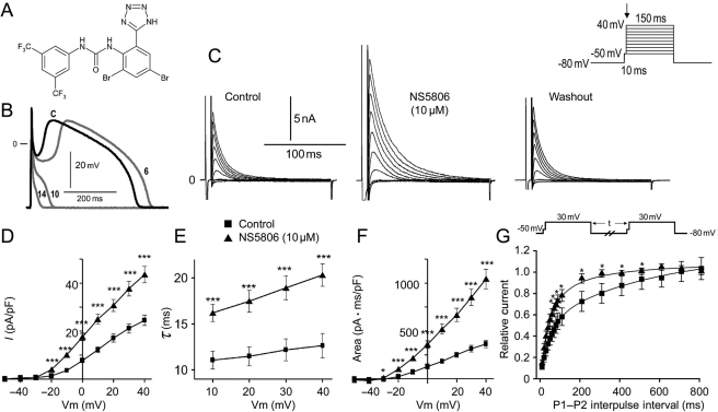
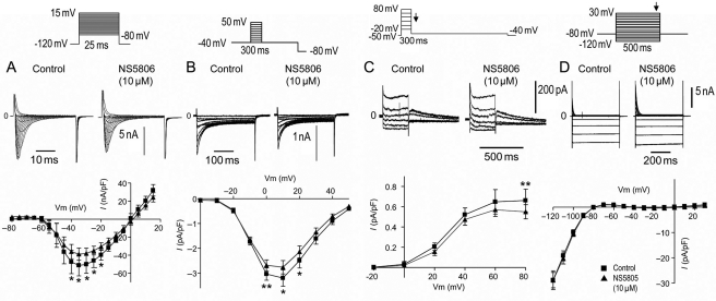
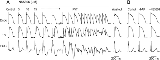
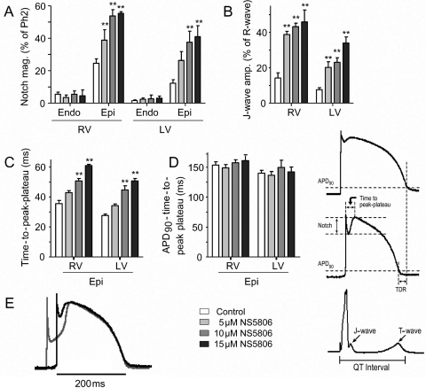
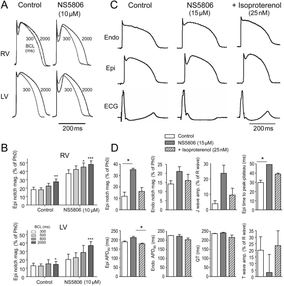
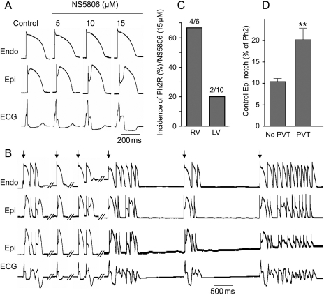
Methods and results: Isolated canine ventricular myocytes and coronary-perfused wedge preparations were used. Whole-cell patch-clamp studies showed that NS5806 (10 microM) increased peak I(to) at +40 mV by 79 +/- 4% (24.5 +/- 2.2 to 43.6 +/- 3.4 pA/pF, n = 7) and slowed the time constant of inactivation from 12.6 +/- 3.2 to 20.3 +/- 2.9 ms (n = 7). The total charge carried by I(to) increased by 186% (from 363.9 +/- 40.0 to 1042.0 +/- 103.5 pA x ms/pF, n = 7). In ventricular wedge preparations, NS5806 increased phase 1 and notch amplitude of the action potential in the epicardium, but not in the endocardium, and accentuated the ECG J-wave, leading to the development of phase 2 re-entry and polymorphic ventricular tachycardia (n = 9). Although sodium and calcium channel blockers are capable of inducing BrS only in right ventricular (RV) wedge preparations, the I(to) activator was able to induce the phenotype in wedges from both ventricles. NS5806 induced BrS in 4/6 right and 2/10 left ventricular wedge preparations.

Conclusion: The I(to) activator NS5806 recapitulates the electrographic and arrhythmic manifestation of BrS, providing evidence in support of its pivotal role in the genesis of the disease. Our findings also suggest that a genetic defect leading to a gain of function of I(to) could explain variants of BrS, in which ST-segment elevation or J-waves are evident in both right and left ECG leads.


Figures

Figure 1
Figure 1
(A) Chemical structure of the diphenylurea 1-(3,5-bis-trifluoromethyl-phenyl)-3-[2,4-dibromo-6-(1H-tetrazol-5-yl)-phenyl]-urea compound, NS5806. (B) Action potentials recorded from a canine left ventricular (LV) mid-myocardial myocyte before (black) and at 6, 10, and 14 s after 10 µM NS5806 (grey). Basic cycle length (BCL), 1 s. Representative of n = 5. (C) Representative Ito recorded from isolated LV mid-myocardial myocytes in the absence and presence of 10 µM NS5806, n = 7. (D) Current–voltage (IV) relation of peak Ito before and after 10 µM NS5806. (E) Time constant of decay of Ito before and after 10 µM NS5806. (F) Graph showing area under the curve, reflecting the total charge carried by Ito. (G) Ito recovery from inactivation using a two-pulse protocol. Statistical significance was evaluated by paired t-test.
Figure 2
Figure 2
Effect of NS5806 on INa and ICaL recorded for isolated canine left ventricular (LV) mid-myocardial cells and on IKr in endocardial LV myocytes. (A) Representative INa traces and average IV relation recorded before and after 10 µM NS5806 (n = 4). (B) ICaL was elicited by the shown protocol preceded by five pre-pulses (from −80 to +20 mV for 200 ms) to ensure constant load of the sarcoplasmic reticulum. Representative ICaL traces and average IV relations recorded before and after 10 µM NS5806 (n = 6). (C) Representative IKr and IKr tail currents measured at −35 mV as a function of voltage at the preceding voltage step before and after the application of 10 µM NS5806 (n = 7). (D) Representative IK1 recordings in mid-myocardial cells (n = 8) and the current–voltage relationship at the end of the step protocol before and after the application of 10 µM NS5806. Statistical significance was evaluated by paired t-test.
Figure 3
Figure 3
Effect of NS5806 to induce the electrocardiographic and arrhythmic manifestation of Brugada syndrome in a canine right ventricular wedge preparation paced from the endocardial surface at a basic cycle length of 2 s. (A) Endocardium and epicardium action potentials and ECG were recorded before and after 5, 10, and 15 µM NS5806, as well as after 30 min of washout. About 15 µM NS5806 induce polymorphic ventricular tachycardia (PVT). Representative of n = 4. (B) About 2 mM 4-aminopyrodine (4-AP) blocked Ito and prevented 10 µM NS5806 from increasing the epicardial notch in a left ventricular wedge. Representative of n = 4.
Figure 4
Figure 4
Effect of NS5806 on action potential (AP) parameters recorded from canine right (RV) and left ventricular (LV) wedge preparations paced at a basic cycle length of 2 s. Composite data of the effect of NS5806 (0–15 µM) on: (A) AP notch magnitude as percentage of the phase 2 amplitude. (B) J-wave amplitude as percentage of the R-wave amplitude. (C) Time-to-peak-plateau in epicardial APs. (D) Repolarization time measured as action potential duration (APD90) minus time-to-peak-plateau. (E) Superimposed LV epicardial APs recorded in control (black trace) and after 15 µM NS5806 (grey trace). The APs were superimposed at the time of peak of phase 2 (n = 5–8).
Figure 5
Figure 5
Effect of NS5806 (0–15 µM) on action potential duration (APD90), QT interval, and transmural dispersion of repolarization (TDR) in canine right (RV) and left (LV) ventricular wedge preparations. BCL, 2 s. (A) APD90; (B) QT interval; (C) epicardial ADP90 plotted as a function of epicardial notch magnitude. (D) TDR calculated as the time interval between repolarization of the endocardial and epicardial action potentials measured at 90%. For the RV preparations, TDR was measured just before and after the dome was lost in the presence of 15 µM NS5806. n = 5–8.
Figure 6
Figure 6
The effect of NS5806 (10 µM) at different pacing rates on the epicardial notch magnitude. (A) Representative action potentials (APs) recorded at increasing basic cycle lengths from 300 to 2000 ms from canine right (RV) and left (LV) ventricular wedge preparations. (B) The epicardial notch magnitude is plotted as percentage of phase 0 amplitude (n = 4–6). (C) Effect of isoproterenol (25 nM) to reverse the effect of NS5806 (15 µM) in a left ventricular wedge preparation. Representative of n = 3. (D) Mean data showing effect of NS5806 and NS5806+iso on various electrophysiological parameters. Results were compared by Friedman's test and Dunn's post-test.
Figure 7
Figure 7
Effect of NS5806 to induce the electrocardiographic and arrhythmic manifestation of Brugada syndrome in a canine left ventricular (LV) wedge preparation paced at a basic cycle length of 2 s. (A) Representative recordings of action potentials and corresponding ECG before and after NS5806 (0–15 µM). (B) The same preparation following 35 min exposure to 15 µM NS5806, showing development of polymorphic ventricular tachycardia (PVT). The arrows indicate paced beats. (C) Incidence of PVT in the presence of 15 µM NS5806 in right ventricular (RV) wedge vs. LV wedge preparations. (D) Size of the epicardial notch in control in preparations exhibiting PVT (n = 6) vs. preparations without PVT (n = 5). Results from RV and LV wedge preparations are pooled.

Comment in

References

    1. Antzelevitch C. Brugada syndrome. Pacing Clin Electrophysiol. 2006;29:1130–1159. - PMC - PubMed
    1. Brugada P, Brugada J. Right bundle branch block, persistent ST segment elevation and sudden cardiac death: a distinct clinical and electrocardiographic syndrome. A multicenter report. J Am Coll Cardiol. 1992;20:1391–1396. - PubMed
    1. Horigome H, Shigeta O, Kuga K, Isobe T, Sakakibara Y, Yamaguchi I, et al. Ventricular fibrillation during anesthesia in association with J waves in the left precordial leads in a child with coarctation of the aorta. J Electrocardiol. 2003;36:339–343. - PubMed
    1. Ogawa M, Kumagai K, Yamanouchi Y, Saku K. Spontaneous onset of ventricular fibrillation in Brugada syndrome with J wave and ST-segment elevation in the inferior leads. Heart Rhythm. 2005;2:97–99. - PubMed
    1. Potet F, Mabo P, Le CG, Probst V, Schott JJ, Airaud F, et al. Novel Brugada SCN5A mutation leading to ST segment elevation in the inferior or the right precordial leads. J Cardiovasc Electrophysiol. 2003;14:200–203. - PubMed

Publication types

MeSH terms

Substances