. 2009 Apr;296(4):F691-9.
doi: 10.1152/ajprenal.90623.2008. Epub 2008 Dec 10.

The Na+-Pi cotransporter PiT-2 (SLC20A2) is expressed in the apical membrane of rat renal proximal tubules and regulated by dietary Pi

Affiliations

The Na+-Pi cotransporter PiT-2 (SLC20A2) is expressed in the apical membrane of rat renal proximal tubules and regulated by dietary Pi

Ricardo Villa-Bellosta et al. Am J Physiol Renal Physiol. 2009 Apr.

Abstract

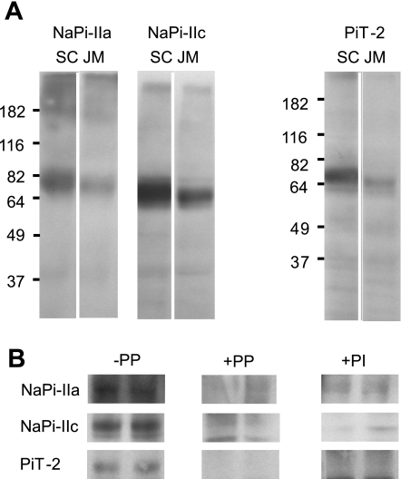
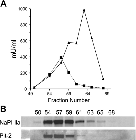
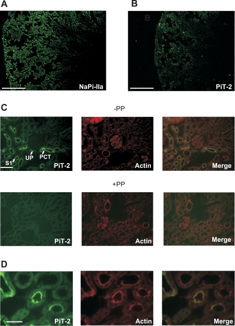
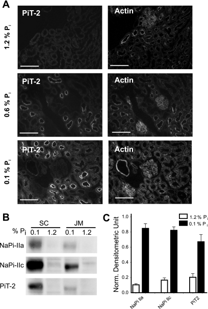
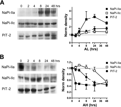
The principal mediators of renal phosphate (P(i)) reabsorption are the SLC34 family proteins NaPi-IIa and NaPi-IIc, localized to the proximal tubule (PT) apical membrane. Their abundance is regulated by circulatory factors and dietary P(i). Although their physiological importance has been confirmed in knockout animal studies, significant P(i) reabsorptive capacity remains, which suggests the involvement of other secondary-active P(i) transporters along the nephron. Here we show that a member of the SLC20 gene family (PiT-2) is localized to the brush-border membrane (BBM) of the PT epithelia and that its abundance, confirmed by Western blot and immunohistochemistry of rat kidney slices, is regulated by dietary P(i). In rats treated chronically on a high-P(i) (1.2%) diet, there was a marked decrease in the apparent abundance of PiT-2 protein in kidney slices compared with those from rats kept on a chronic low-P(i) (0.1%) diet. In Western blots of BBM from rats that were switched from a chronic low- to high-P(i) diet, NaPi-IIa showed rapid downregulation after 2 h; PiT-2 was also significantly downregulated at 24 h and NaPi-IIc after 48 h. For the converse dietary regime, NaPi-IIa showed adaptation within 8 h, whereas PiT-2 and NaPi-IIc showed a slower adaptive trend. Our findings suggest that PiT-2, until now considered as a ubiquitously expressed P(i) housekeeping transporter, is a novel mediator of P(i) reabsorption in the PT under conditions of acute P(i) deprivation, but with a different adaptive time course from NaPi-IIa and NaPi-IIc.


Figures

Fig. 1.
Fig. 1.
Detection of Na+-coupled Pi cotransporter (NaPi)-IIa, NaPi-IIc, and PiT-2 proteins in kidney brush-border membrane (BBM) preparations by immunoblotting. A: Western blots of BBM from superficial cortical (SC) and juxtamedullary (JM) regions for NaPi-IIa, NaPi-IIc, and PiT-2 from the same animal. In this and other Western blots, 34 μg protein were loaded per lane. NaPi-IIa affinity antibody was used at 1:3,000 dilution in PBS, NaPi-IIc at 1:5,000, and PiT-2 at 1:3,000. B: specificity of the antibodies. −PP, immunoreactive bands in control kidney cortex BBM proteins; +PP, blocking of antibodies with the immunogenic peptide. Affinity purified antibodies were incubated with 50 μg/ml peptide for 5 min at room temperature before the Western blot assay. +PI, blots obtained with preimmune sera of the corresponding animals.
Fig. 2.
Fig. 2.
Free-flow electrophoresis. Free-flow electrophoretic separation of rat kidney cortical BBM and basolateral membranes. Single fractions were analyzed for enzyme activity in leucine aminopeptidase N (▪) and Na+-K+-ATPase (▴) (A) and probed by Western blot analysis for NaPi-IIa and PiT-2 (B).
Fig. 3.
Fig. 3.
PiT-2 immunohistochemistry for kidney slices from one animal fed on a chronic low-Pi (0.1%) diet. A: immunostaining with PiT-2 antibody indicates expression of PiT-2, particularly in cortical areas. Scale bar: 750 μm. B: immunostaining with NaPi-IIa antibody of material from same animal shows more widespread staining in both cortical and JM regions. Scale bar: 750 μm. C: immunostaining with PiT-2 antibody of kidney slice at higher magnification shows strong staining in the early part of the proximal tubule, including urinary pole (UP), proximal convoluted tubule (PCT), and S1 segment of PT (S1). Scale bar: 375 μm. Second row shows images with peptide protection (PP). D: higher magnification of S1 segment staining for PiT-2 (left), actin (center), and merged images (right). Scale bar: 75 μm.
Fig. 4.
Fig. 4.
Response to chronic dietary conditions. A: immunohistochemistry of kidney slice stained for PiT-2 (left) and for actin (right). Top: chronic high-Pi (1.2%) diet; center: normal-Pi (0.6%) diet; bottom: chronic low-Pi (0.1%) diet. Scale bar: 150 μm. B: Western blots from SC and JM BBM using material from animals fed on chronic high-Pi and chronic low-Pi diets. C: quantitation of Western blots for NaPi-IIa, NaPi-IIc, and PiT-2 indicates that all three transporters respond similarly to chronic changes in Pi concentration in the diet (n = 5 experiments). The downregulation, based on densitometric analysis, of material from rats treated on low-Pi and high-Pi diet is 88% for NaPi-IIa (P < 0.0001), 84% for NaPi-IIc (P < 0.0001), and 70% for PiT-2 (P = 0.0023). Normalized densitometric units were calculated by subtracting the background from the signal and corrected using the actin signal.
Fig. 5.
Fig. 5.
Acute adaptation of NaPi-IIa, NaPi-IIc, and PiT-2 to dietary conditions. A: changes from chronic high-Pi diet to acute low-Pi diet (AL). Western blots from representative animals are shown for NaPi-IIa, NaPi-IIc, and PiT-2. In each case, densitometric quantitation of acute changes shows the mean of measurements on material taken from 3 animals killed at the indicated time point. Whereas NaPi-IIa is upregulated within 4 h of dietary change to low-Pi diet, NaPi-IIc and PiT-2 require 8 h to increase their abundance at the plasma membrane. B: changes from chronic low-Pi diet to acute high-Pi diet (AH) as in A. NaPi-IIa is eliminated from the BBM within 2 h, whereas the reduction in NaPi-IIc expression is only clearly seen at 48 h, and PiT-2 at 8 h.

Comment in

  • PiT-2 coming out of the pits.
    Moe OW. Moe OW. Am J Physiol Renal Physiol. 2009 Apr;296(4):F689-90. doi: 10.1152/ajprenal.00007.2009. Epub 2009 Feb 4. Am J Physiol Renal Physiol. 2009. PMID: 19193727 No abstract available.

References

    1. Bacconi A, Virkki LV, Biber J, Murer H, Forster IC. Renouncing electrogenicity is not free of charge: switching on electrogenicity in a Na+-coupled phosphate cotransporter. Proc Natl Acad Sci USA 102: 12606–12611, 2005. - PMC - PubMed
    1. Bai L, Collins JF, Ghishan FK. Cloning and characterization of a type III Na-dependent phosphate cotransporter from mouse intestine. Am J Physiol Cell Physiol 279: C1135–C1143, 2000. - PubMed
    1. Beck L, Karaplis AC, Amizuka N, Hewson AS, Ozawa H, Tenenhouse HS. Targeted inactivation of Npt2 in mice leads to severe renal phosphate wasting, hypercalciuria, and skeletal abnormalities. Proc Natl Acad Sci USA 95: 5372–5377, 1998. - PMC - PubMed
    1. Berndt T, Kumar R. Phosphatonins and the regulation of phosphate homeostasis. Annu Rev Physiol 69: 341–359, 2007. - PubMed
    1. Berndt TJ, Kumar R. Clinical disturbances of phosphate homeostasis. In: Seldin and Giebisch's The Kidney, edited by Alpern RJ and Hebert SC. New York, NY: Academic, 2008, p. 1989–2006.

Publication types

Substances