. 2009 Feb;37(2):602-10.
doi: 10.1093/nar/gkn922. Epub 2008 Dec 10.

The C-terminal fragment of the ribosomal P protein complexed to trichosanthin reveals the interaction between the ribosome-inactivating protein and the ribosome

Affiliations

The C-terminal fragment of the ribosomal P protein complexed to trichosanthin reveals the interaction between the ribosome-inactivating protein and the ribosome

Priscilla Hiu-Mei Too et al. Nucleic Acids Res. 2009 Feb.

Abstract

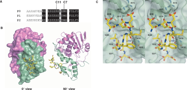
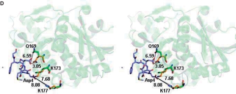
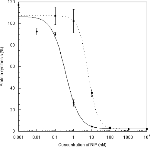
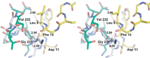
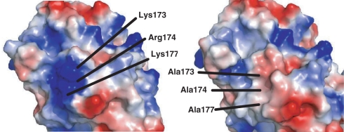
Ribosome-inactivating proteins (RIPs) inhibit protein synthesis by enzymatically depurinating a specific adenine residue at the sarcin-ricin loop of the 28S rRNA, which thereby prevents the binding of elongation factors to the GTPase activation centre of the ribosome. Here, we present the 2.2 A crystal structure of trichosanthin (TCS) complexed to the peptide SDDDMGFGLFD, which corresponds to the conserved C-terminal elongation factor binding domain of the ribosomal P protein. The N-terminal region of this peptide interacts with Lys173, Arg174 and Lys177 in TCS, while the C-terminal region is inserted into a hydrophobic pocket. The interaction with the P protein contributes to the ribosome-inactivating activity of TCS. This 11-mer C-terminal P peptide can be docked with selected important plant and bacterial RIPs, indicating that a similar interaction may also occur with other RIPs.


Figures

Figure 1.
Figure 1.
(A) Sequence alignment of the C-terminal residues of mammalian P proteins. The last 17 aa of the P proteins are nearly identical. Positions of the last 11 and 7 aa of the P proteins are marked. (B) The c11-P motif (SDDDMGFGLFD) (yellow) of eukaryotic stalk proteins binds to a pocket in the C-terminal domain of TCS. The N-terminal and the C-terminal domains of TCS are colour-coded violet and green, respectively. (C) A stereo diagram showing the interaction between c11-P (yellow) and TCS (green). Intermolecular hydrogen bonds and salt-bridges are indicated as dashed lines. Residue numbers of TCS and c11-P are in black and blue, respectively. (D) A stereo diagram showing the movement of TCS side chains upon interacting with c11-P. Compared to the wild-type TCS (green), the side chain of Gln169 in TCS–c11-P (orange) flips toward Asp2 of c11-P and forms a hydrogen bond with that residue; the side chain of Lys173 is located between OD1 and OD2 of Asp4; the side chain of Lys177 moves from 8.08 Å to 7.68 Å toward Asp4 of the peptide.
Figure 1.
Figure 1.
(A) Sequence alignment of the C-terminal residues of mammalian P proteins. The last 17 aa of the P proteins are nearly identical. Positions of the last 11 and 7 aa of the P proteins are marked. (B) The c11-P motif (SDDDMGFGLFD) (yellow) of eukaryotic stalk proteins binds to a pocket in the C-terminal domain of TCS. The N-terminal and the C-terminal domains of TCS are colour-coded violet and green, respectively. (C) A stereo diagram showing the interaction between c11-P (yellow) and TCS (green). Intermolecular hydrogen bonds and salt-bridges are indicated as dashed lines. Residue numbers of TCS and c11-P are in black and blue, respectively. (D) A stereo diagram showing the movement of TCS side chains upon interacting with c11-P. Compared to the wild-type TCS (green), the side chain of Gln169 in TCS–c11-P (orange) flips toward Asp2 of c11-P and forms a hydrogen bond with that residue; the side chain of Lys173 is located between OD1 and OD2 of Asp4; the side chain of Lys177 moves from 8.08 Å to 7.68 Å toward Asp4 of the peptide.
Figure 2.
Figure 2.
Assay for the inhibition of protein synthesis. The ability of wild-type and [V232K/N236D]TCS to inhibit the incorporation of [3H]-leucine into the proteins of the nuclease-untreated rabbit reticulocyte lysate was assayed as described (4). Each point denotes the average of an assay done in triplicate. The IC50, the concentration of RIP required to achieve 50% inhibition, was determined by fitting the data to a four-parameter logistic equation. The values of IC50 for wild-type (filled circle) and [V232K/N236D]TCS (filled square) were 0.37 and 6.28 nM, respectively.
Figure 3.
Figure 3.
Interaction with the ribosomal stalk for full activity of TCS. (A) Rat 80S ribosome was loaded onto NHS-Sepharose coupled with TCS, [V232K/N236D]TCS or [K173A/R174A/K177A]TCS; the elutants were detected with a Western blot using an anti-P antibody. Ribosomal protein P0 was pull-down by the wild-type TCS (lane 2) but not by TCS variants (lane 3 and 4). (B) Disuccinimidyl suberate (DSS) was used to induce cross-linking between TCS and the ribosome. A protein band at 66 kDa, corresponding to the size of the TCS–P0 complex, was detected by both anti-P and anti-TCS antibodies when the ribosome was cross-linked with wild-type TCS (lane 4) but not with the variants (lane 7 and 10) or negative controls. (C) Depurinated rRNA was hydrolysed by acidic aniline; the 450-bp R-fragments (indicated by an arrow) indicate specific depurination of the 28S rRNA at A4324. Control lanes, in which RNA samples were not treated with aniline, are labeled with the ‘–’ marks. Only wild-type TCS had specific N-glycosidase activity.
Figure 4.
Figure 4.
Stereo image showing the superimposition of the TCS–c-11P complex and [V232K/N236D]TCS. Note that the side chain of Lys232 is flipped out to the solvent accessible surface. The movement of Lys232-induced changes in the conformation of neighbouring residues; remarkably, the Cα atoms of Ala230 and Gly231 are shifted by 1.98 and 3.43 Å, respectively. The three hydrogen bond interactions that were interrupted by the mutations are indicated. TCS–c-11P complex is coloured in green, and [V232K/N236D]TCS is coloured in white.
Figure 5.
Figure 5.
Electrostatic surface representation of (A) Wild-type TCS and (B) [K173A/R174A/K177A]TCS, indicating the loss of a patch of surface positive charges in the latter. Positive charge is shown in blue, and negative charge is in red.
Figure 6.
Figure 6.
Stereo images of the molecular docking of c11-P peptide to RTA, SO6 and SLT-1A. (A) RTA, (B) SO6 and (C) SLT-1A. c11-P is shown in ball and stick representation and in green colour. Potential hydrogen bond interactions between the RIPs and the c-11 peptide are indicated.

References

    1. Tchorzewski M. The acidic ribosomal P proteins. Int. J. Biochem. Cell Biol. 2002;34:911–915. - PubMed
    1. Helgstrand M, Mandava CS, Mulder FAA, Liljas A, Sanyal S, Akke M. The ribosomal stalk binds to translation factors IF2, EF-Tu, EF-G and RF3 via a conserved region of the L12 C-terminal domain. J. Mol. Biol. 2007;365:468–479. - PubMed
    1. Shaw PC, Lee KM, Wong KB. Recent advances in trichosanthin, a ribosome-inactivating protein with multiple pharmacological properties. Toxicon. 2005;45:683–689. - PubMed
    1. Chan DSB, Chu LO, Lee KM, Too PHM, Ma KW, Sze KH, Zhu G, Shaw PC, Wong KB. Interaction between trichosanthin, a ribosome-inactivating protein, and the ribosomal stalk protein P2 by chemical shift perturbation and mutagenesis analyses. Nucleic Acids Res. 2007;35:1660–1672. - PMC - PubMed
    1. McCluskey AJ, Poon GMK, Bolewska-Pedyczak E, Srikumar T, Jeram SM, Raught B, Gariépy J. The catalytic subunit of Shiga-like toxin 1 interacts with ribosomal stalk proteins and is inhibited by their conserved C-terminal domain. J. Mol. Biol. 2008;378:375–386. - PubMed

Publication types