. 2009 Mar;83(5):2237-45.
doi: 10.1128/JVI.01699-08. Epub 2008 Dec 10.

Reevaluating the CD8 T-cell response to herpes simplex virus type 1: involvement of CD8 T cells reactive to subdominant epitopes

Affiliations

Reevaluating the CD8 T-cell response to herpes simplex virus type 1: involvement of CD8 T cells reactive to subdominant epitopes

Brian S Sheridan et al. J Virol. 2009 Mar.

Abstract

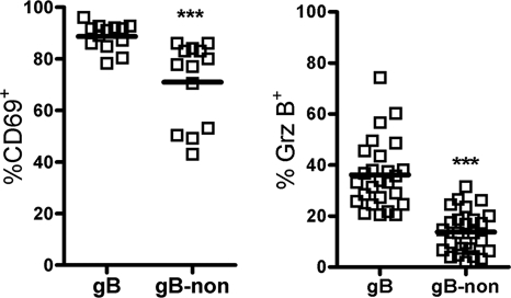
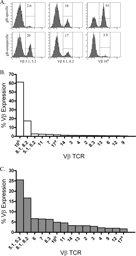
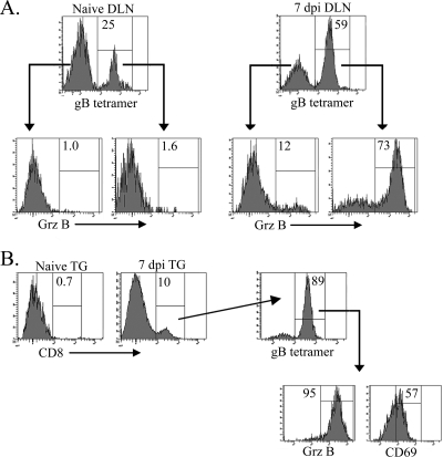
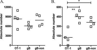
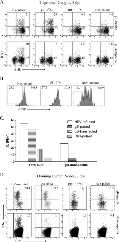
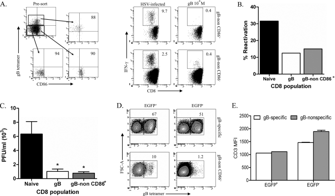
In C57BL/6 (B6) mice, most herpes simplex virus (HSV)-specific CD8 T cells recognize a strongly immunodominant epitope on glycoprotein B (gB498) and can inhibit HSV type 1 (HSV-1) reactivation from latency in trigeminal ganglia (TG). However, half of the CD8 T cells retained in latently infected TG of B6 mice are not gB498 specific and have been largely ignored. The following observations from our current study indicate that these gB498-nonspecific CD8 T cells are HSV specific and may contribute to the control of HSV-1 latency. First, following corneal infection, OVA257-specific OT-1 CD8 T cells do not infiltrate the infected TG unless mice are simultaneously immunized with OVA257 peptide, and then they are not retained. Second, 30% of CD8 T cells in acutely infected TG that produce gamma interferon in response to HSV-1 stimulation directly ex vivo are gB498 nonspecific, and these cells maintain an activation phenotype during viral latency. Finally, gB498-nonspecific CD8 T cells are expanded in ex vivo cultures of latently infected TG and inhibit HSV-1 reactivation from latency in the absence of gB498-specific CD8 T cells. We conclude that many of the CD8 T cells that infiltrate and are retained in infected TG are HSV specific and potentially contribute to maintenance of HSV-1 latency. Identification of the viral proteins recognized by these cells will contribute to a better understanding of the dynamics of HSV-1 latency.


Figures

FIG. 1.
FIG. 1.
The gB498-nonspecific CD8 T-cell population within infected TG is activated and undergoes proliferation. (A) TG were harvested at indicated dpi and stained with gB498 tetramers for quantification of gB498-specific and gB498-nonspecific CD8 T cells. The inset shows staining of CD8 T cells in TG at 8 dpi and illustrates the comparable sizes of the gB498-specific and gB498-nonspecific CD8 T-cell populations observed at all times beyond 8 dpi. Data are presented as the mean absolute number of each CD8 T-cell population in individual TG ± standard error of the mean. (B) HSV-1-infected mice received 1 mg BrdU i.v. 4 h prior to TG excision, and in situ proliferation of CD8 T-cell populations was assessed on 7 dpi by intracellular staining for incorporated BrdU and flow cytometry. Representative dot plots are gated on either gB498-specific or gB498-nonspecific CD8 T cells as demonstrated in the inset to panel A, gates are based on appropriate isotype controls, and numbers within dot plots represent the percentage of cells within the positive gate. Data from individual mice are represented as indicated (mean ± standard error of the mean). (C) Phenotypic characterization of CD8 T-cell populations within the TG at 7 dpi. Representative histograms are gated on either gB498-specific or gB498-nonspecific CD8 T cells as illustrated in the inset to panel A, gates are based on appropriate isotype controls, and numbers within histograms correspond to the percentage of cells within the positive gate. Data from individual mice are presented in the scatter plot as percent expression of indicated marker within the gB498-specific and gB498-nonspecific CD8 T-cell populations (solid line = mean). ***, P < 0.001; **, P < 0.01 (Student's t test).
FIG. 2.
FIG. 2.
Maintained activation of gB498-nonspecific CD8 T cells within the latent ganglia. B6 mice were allowed to establish latency (>34 days) prior to excision of the TG. Single-cell suspensions were stained for expression of CD8, for expression of CD69, with gB498 tetramers, and intracellularly for grz B. Data from individual mice are presented in the scatter plot as percent expression of the indicated marker within the gB498-specific and gB498-nonspecific CD8 T-cell populations (solid line = mean). ***, P < 0.001 (Student's t test).
FIG. 3.
FIG. 3.
The gB498-nonspecific CD8 T-cell population displays a more diverse Vβ TCR usage. TG were harvested at 8 dpi, pooled, and stained with gB498 tetramers, for CD8, and for TCR Vβ utilization. (A) Representative histograms illustrate utilization of three prominent Vβ (Vβ 5.1, 5.2, Vβ 8.1, 8.2, and Vβ 10b) TCR segments by gB498 tetramer-positive (gB498-specific) and gB498 tetramer-negative (gB498-nonspecific) CD8 T cells. Numbers within histograms correspond to the percentage of cells within the positive gate. (B and C) Data are presented graphically as the percent expression of indicated Vβ TCR among gB498-specific CD8 T cells (B) or gB498-nonspecific CD8 T cells (C).
FIG. 4.
FIG. 4.
Bystander activation does not appear to account for CD8 T-cell infiltration into the infected TG. CD8 T cells were isolated from spleens of noninfected gB-T1.1 and OT-1 mice and transferred into CD8−/− mice 1 day prior to mock infection or infection with HSV-1. Both the DLN (A) and TG (B) were excised at 7 dpi, and gB498-specific CD8 T cells were quantified using gB498 tetramers. Mock-infected recipient mice are labeled naïve. Parent histograms from representative mice are gated on CD8 T cells (A) or CD45 cells (B) with the gating strategy depicted. Numbers within histograms correspond to the percentage of cells within the positive gate.
FIG. 5.
FIG. 5.
Known HSV-1-nonspecific CD8 T cells are not retained in the latent ganglia. CD45.2+ OT-1 CD8 T cells were isolated from naïve spleens, and 105 were transferred into B6.SJL (CD45.1) mice 1 day prior to simultaneous infection with HSV-1 and DC-OVA257 immunization. TG of these mice were harvested 8 (A) and 40 (B) days later for characterization of the infiltrating populations. Host and donor populations were distinguished based on congenic CD45 markers, and host populations are further delineated by recognition with gB498 mulitmers. The absolute number of each CD8 T-cell population in individual TG is represented graphically, with a solid line indicating the mean. **, P < 0.01; *, P < 0.05 (one-way analysis of variance with Bonferroni post hoc t test).
FIG. 6.
FIG. 6.
A proportion of gB498-nonspecific CD8 T cells recognize other HSV-1 epitopes. (A) B6 mice were infected with HSV-1 and given 1 mg BrdU i.p. daily beginning at 5 dpi. Pooled TG were harvested at indicated dpi, sorted into either gB498-specific or gB498-nonspecific CD8 T cells, and then stimulated with the indicated targets for 6 h in the presence of brefeldin A. Cell suspensions were then stained for surface expression of CD45 and CD8, permeabilized, and stained intracellularly for IFN-γ and BrdU. Representative dot plots show the percentage of cells within the respective quadrants. (B) B6 mice were infected with HSV-1, and pooled TG were harvested at 8 dpi. A pure population of gB498-nonspecific CD8 T cells was obtained by sorting, labeled with 1.0 μM CFSE, and stimulated with the indicated targets for 3 days at 37°C with 5% CO2. Cell suspensions were collected and stained for surface expression of CD45 and CD8 and analyzed for CFSE dilution. Histograms show the percentage of cells within the respective quadrant on the left and the CFSE MFI of the entire CD8 population on the right. (C) Pooled TG obtained at 8 dpi were dispersed into single cell suspensions, stained for 1 h with PE- conjugated gB498 tetramers, and stimulated for 6 h with syngeneic targets that were HSV-1 infected, gB498 pulsed, RR1822 pulsed, or transfected to express full-length gB in the presence of brefeldin A. The cells were then permeabilized, fixed, and stained for intracellular IFN-γ. (D) DLN cells obtained at 7 dpi were stained with gB498 tetramers; sorted into tetramer-positive (gB498-specific) and tetramer-negative (gB498-nonspecific) populations; stimulated for 6 h with HSV-1 infected, gB498-pulsed, RR1822-pulsed, or unmanipulated targets in the presence of brefeldin A; stained for intracellular IFN-γ; and analyzed by flow cytometry. Representative dot plots show the percentage of sorted CD8 T cells positive for intracellular IFN-γ.
FIG. 7.
FIG. 7.
gB498-nonspecific CD8 T cells are capable of preventing reactivation and impairing viral spread following TG explant. (A) Pooled spleens from mice at 8 dpi were stained with anti-CD8, anti-CD86, and gB498 tetramer. Cells were then sorted into three populations: (i) gB498-specific CD8 T cells, (ii) gB498-nonspecific CD86+ CD8 T cells, and (ii) gB498-nonspecific CD86 CD8 T cells. Pre- and postsort dot plots and purities are shown. Sorted populations were then stimulated for 6 h in the presence of brefeldin A with the indicated targets. Following stimulation, cell suspensions were stained for surface expression of CD45 and CD8 and then intracellularly for IFN-γ. Representative dot plots are shown with the percentage of IFN-γ-producing cells. (B) HSV-1 pICP0-EGFP latently infected TG cultures depleted of CD45-expressing cells via complement fixation were plated in 0.2 TG equivalent in 48-well plates. Cells sorted as described above were added to the cultures, and the cultures were monitored for reactivation by spread of fluorescence from neurons to surrounding fibroblasts and by detection of infectious virus in culture supernatants in a viral plaque assay with equivalent results. The graph depicts cumulative reactivation frequencies during 11 days in culture. (C and D) After 11 days in culture, infectious virus in supernatants of individual cultures that exhibited HSV-1 reactivation (based on EGFP spread) was quantified (C) and cells were collected from pooled wells in which the virus reactivated (EGFP+) and from those in which the virus did not reactivate (EGFP) (D). Cells were stained with anti-CD8, anti-CD45, anti-CD3, and gB498 tetramers. Representative contour plots show the CD8 T cells isolated from pooled wells which received either gB498-specific or gB498-nonspecific CD86+ CD8 T cells. Numbers within contour plots represent the percentage of CD8 T cells that bound gB498 tetramer. (E) Quantification of CD3 MFI for pooled wells as described above is represented graphically as the mean CD3 MFI (± standard error of the mean). *, P < 0.05 (one-way analysis of variance with a Bonferroni post hoc t test).

Similar articles

Cited by

References

    1. Allan, R. S., J. Waithman, S. Bedoui, C. M. Jones, J. A. Villadangos, Y. Zhan, A. M. Lew, K. Shortman, W. R. Heath, and F. R. Carbone. 2006. Migratory dendritic cells transfer antigen to a lymph node-resident dendritic cell population for efficient CTL priming. Immunity 25153-162. - PubMed
    1. Baron, C., M. C. Meunier, E. Caron, C. Cote, M. J. Cameron, D. J. Kelvin, R. LeBlanc, V. Rineau, and C. Perreault. 2006. Asynchronous differentiation of CD8 T cells that recognize dominant and cryptic antigens. J. Immunol. 1778466-8475. - PubMed
    1. Coles, R. M., S. N. Mueller, W. R. Heath, F. R. Carbone, and A. G. Brooks. 2002. Progression of armed CTL from draining lymph node to spleen shortly after localized infection with herpes simplex virus 1. J. Immunol. 168834-838. - PubMed
    1. Coulombe, M., and R. G. Gill. 1996. T lymphocyte indifference to extrathymic islet allografts. J. Immunol. 1561998-2003. - PubMed
    1. Decman, V., M. L. Freeman, P. R. Kinchington, and R. L. Hendricks. 2005. Immune control of HSV-1 latency. Viral Immunol. 18466-473. - PubMed

Publication types