. 2009 Feb;83(4):1952-61.
doi: 10.1128/JVI.01578-08. Epub 2008 Dec 10.

Role for plasmacytoid dendritic cells in the immune control of recurrent human herpes simplex virus infection

Affiliations

Role for plasmacytoid dendritic cells in the immune control of recurrent human herpes simplex virus infection

Heather Donaghy et al. J Virol. 2009 Feb.

Abstract

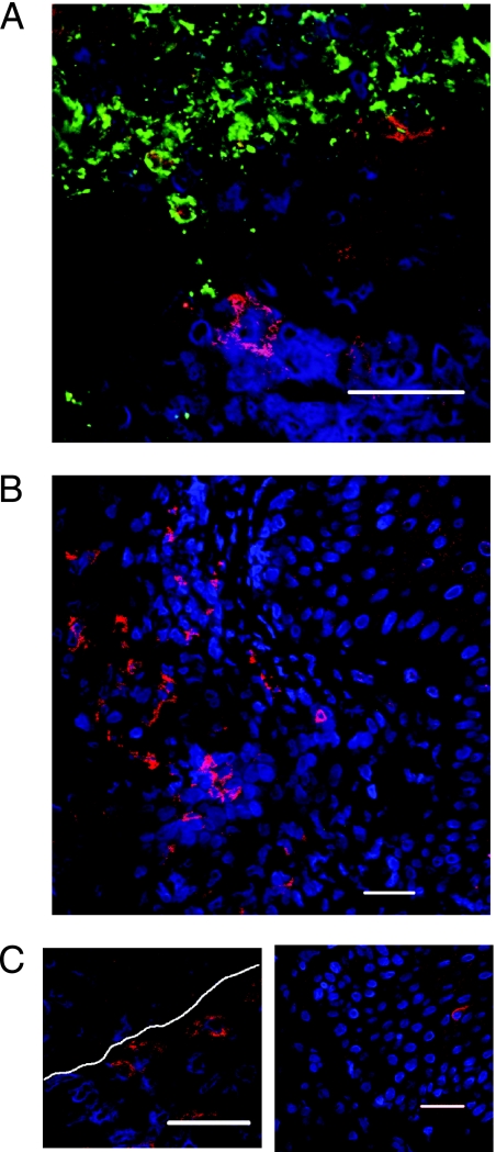
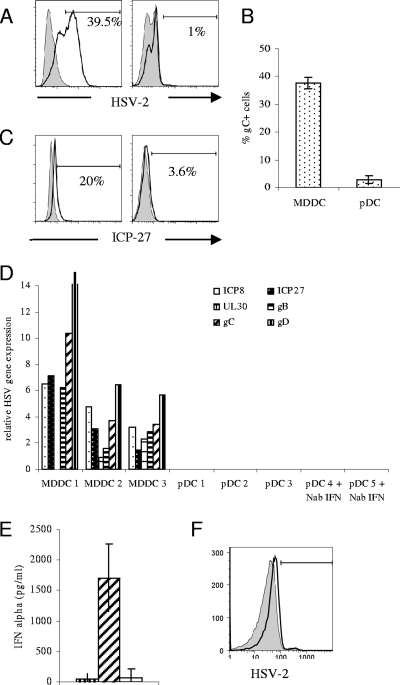
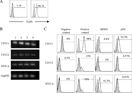
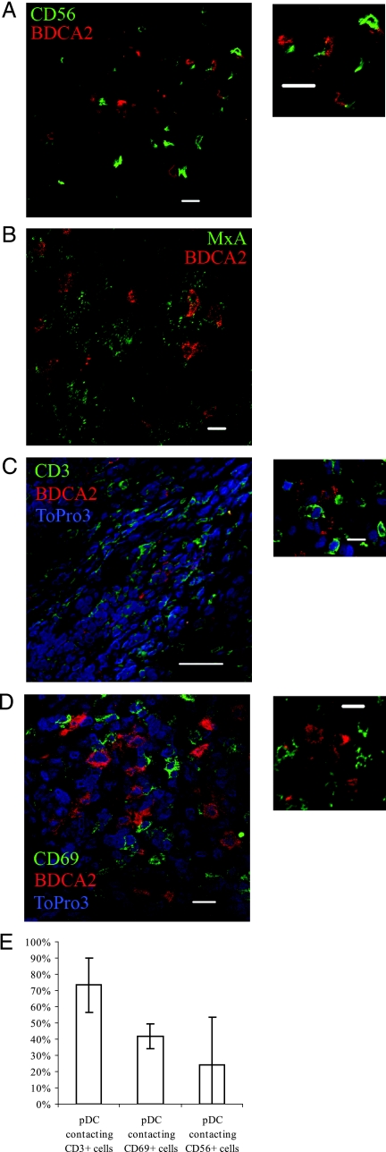
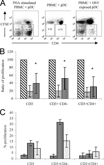
Plasmacytoid dendritic cells (pDC) are an important component of the innate immune response, producing large amounts of alpha interferon in response to viral stimulation in vitro. Under noninflammatory conditions, pDC are not found in the skin and are restricted in location to the blood and lymph nodes. Therefore, their role in mucosal and cutaneous herpes simplex virus (HSV) infection has not been well-defined. In this study we show a role for human pDC in the immune response to HSV infection. First, by confocal microscopy we showed that pDC infiltrate the dermis of recurrent genital herpes simplex lesions at early and late phases, often at the dermo-epidermal junction. We then showed that pDC in vitro are resistant to HSV infection despite expressing the entry receptors CD111, CD112, and HVE-A. Within the lesions, pDC were found closely associated with CD3(+) lymphocytes and NK cells, especially those which were activated (CD69(+)). Furthermore, these HSV-exposed pDC were able to stimulate virus-specific autologous T-lymphocyte proliferation. We conclude from this work that pDC may contribute to the immune control of recurrent herpes virus infection in vivo.


Figures

FIG. 1.
FIG. 1.
Plasmacytoid pDC are detected in recurrent herpes simplex type 2 biopsies. Biopsies of herpes simplex type 2 lesions were stained for BDCA-2 (red), TO-PRO-3 (blue), and HSV-2 (green) and analyzed by confocal microscopy. (A) Biopsy of herpes simplex type 2 lesion stained with BDCA-2, TO-PRO-3, and HSV-2, indicating infiltration of pDC into virally infected lesions. (B) BDCA-2+ cells were commonly observed in the dermis. (C) BDCA-2+ cells were also found at the junction between the dermis and epidermis (left panel) and occasionally in the epidermis (right panel). White dotted lines indicate the dermo-epidermal junction as determined histologically. Bars, 10 μm.
FIG. 2.
FIG. 2.
Plasmacytoid DC exposed to HSV-2 in vitro are resistant to infection. MDDC and pDC were exposed to HSV-2 at a multiplicity of infection of 5. Also, pDC were cultured in the presence of neutralizing antibodies to IFN-α, IFN-β, and IFN-α/β receptor prior to exposure to HSV-2. (A and B) Surface HSV-2 gB expression was measured by flow cytometry 16 h postinfection. Representative data from one individual (A) (left panel, MDDC; right panel, pDC) and mean data from three MDDC and nine pDC donors (B) are shown. Error bars indicate standard deviations. (C) After 6 h of infection, intracellular ICP27 expression was measured by flow cytometry (left panel, MDDC; right panel, pDC). (D) At 16 h postinfection cells were lysed and quantitative PCR of six viral gene transcripts, representing immediate-early proteins (ICP8 and ICP27), early proteins (UL30), and late proteins (gB, gC, and gD) was performed. Expression of viral gene products relative to the housekeeping gene GAPDH are shown for each of three independent experiments. (E) IFN-α production by pDC 16 h postinfection. The first bar indicates mock-infected cells, the second shows results following HSV stimulation, and the third bar indicates ablation of IFN-α following culture in the presence of neutralizing antibodies to IFN-α, IFN-β, and IFN-α/β receptor. IFN-α was measured by ELISA, and the means of three separate experiments are shown. (F) Flow cytometric analysis of HSV-2 expression following culture in the presence of neutralizing antibodies to IFN-α, IFN-β, and IFN-α/β receptor indicates no increase in infectivity following treatment with neutralizing antibody. The gray histogram indicates infection of pDC with HSV (2.5%), and the black open histogram shows infection following addition of neutralizing antibodies to HSV (5%). Results shown are representative of three independent experiments.
FIG. 3.
FIG. 3.
Expression of TLR9 and HSV entry receptors by pDC. TLR9 expression by isolated pDC was measured by flow cytometry (A). Surface (left panel) and intracellular (right panel) staining were performed on freshly isolated pDC by using flow cytometry. Numbers indicate percentages of cells positive for TLR9. (B) PCR was performed using primers for the common HSV entry receptors. Both MDDC and pDC expressed all three HSV receptors. Results from three independent experiments are shown. Lane 1, pDC; lane 2, pDC + HSV; lane 3, MDDC; lane 4, MDDC + HSV. (C) Flow cytometric analysis of expression of HSV entry receptors on different cell types was performed using specific antibodies to CD111 (CK41), CD112 (MP9), and HVE-A (CW10). HeLa cells were used as negative control cells for expression of CD111 and positive controls for CD112 and HVE-A. Jurkat cells were negative controls for CD112 and HVE-A, and HL60 cells were positive controls for CD111. The first panel shows expression in negative control cell lines, the second is positive control expression, the third panel shows levels of expression on MDDC, and the fourth panel shows expression in pDC.
FIG. 4.
FIG. 4.
Plasmacytoid DC interactions in herpes lesions. Biopsies of herpes simplex lesions were stained for BDCA-2 (red), TO-PRO-3 (blue), and (shown in green) for CD56 (A), MxA (B), CD3 (C), or CD69 (D) and analyzed by confocal microscopy. BDCA4+ pDC were found to interact with CD56+ NK cells, CD3+ T cells, and CD69+-activated cells, and they were also found in close proximity to MxA-producing cells. (E) Percentage of pDC contacting CD3, CD69, and CD56 cells in HSV lesions. Means of four fields are indicated. Bars, 10 μm.
FIG. 5.
FIG. 5.
Isolated pDC exposed to HSV-2 stimulate autologous T-lymphocyte proliferation. Isolated pDC were exposed to HSV-2 for 1 h, washed three times, and cultured for a further 16 h. pDC were cultured with autologous CFSE-labeled PBMC for 5 days, and the proliferation induced in CD3+ cells was determined by CFSE dilution by flow cytometry. CD3+ cells were further divided into CD3+ CD4+ and CD3+ CD4 fractions, and proliferation in each population was measured. (A) Representative flow cytometric data from one donor. The numbers in the boxes indicate percentages of CD3+ cells that proliferated. (B) Mean proliferation from seven independent individuals. Data were normalized against the proliferation induced by PHA stimulation (100%). Bars indicate the relative proliferation of autologous PBMC when cocultured with PHA (diagonal stripes), unstimulated pDC (horizontal stripes), and HSV-exposed pDC (vertical stripes). (C) pDC exposed to UV-inactivated HSV stimulate CD3 T-cell proliferation. Bars indicate percentages of CD3+, CD3+ CD4, and CD3+ CD4+ cells in the proliferating gate (i.e., those cells that had a reduction in the expression of CFSE) following exposure to unstimulated pDC (horizontal stripes), HSV-exposed pDC (vertical stripes), and UV-inactivated HSV-exposed pDC (dots). Results are the means of two independent experiments. Error bars indicate standard deviations. *, P < 0.05, determined by paired t test, between unstimulated pDC and HSV-exposed pDC.

References

    1. Agis, H., W. Fureder, H. C. Bankl, M. Kundi, W. R. Sperr, M. Willheim, G. Boltz-Nitulescu, J. H. Butterfield, K. Kishi, K. Lechner, and P. Valent. 1996. Comparative immunophenotypic analysis of human mast cells, blood basophils and monocytes. Immunology 87535-543. - PMC - PubMed
    1. Albers, I., H. Kirchner, and I. Domke-Opitz. 1989. Resistance of human blood monocytes to infection with herpes simplex virus. Virology 169466-469. - PubMed
    1. Allan, R. S., C. M. Smith, G. T. Belz, A. L. van Lint, L. M. Wakim, W. R. Heath, and F. R. Carbone. 2003. Epidermal viral immunity induced by CD8α+ dendritic cells but not by Langerhans cells. Science 3011925-1928. - PubMed
    1. Allan, R. S., J. Waithman, S. Bedoui, C. M. Jones, J. A. Villadangos, Y. Zhan, A. M. Lew, K. Shortman, W. R. Heath, and F. R. Carbone. 2006. Migratory dendritic cells transfer antigen to a lymph node-resident dendritic cell population for efficient CTL priming. Immunity 25153-162. - PubMed
    1. Ashkar, A. A., S. Bauer, W. J. Mitchell, J. Vieira, and K. L. Rosenthal. 2003. Local delivery of CpG oligodeoxynucleotides induces rapid changes in the genital mucosa and inhibits replication, but not entry, of herpes simplex virus type 2. J. Virol. 778948-8956. - PMC - PubMed

MeSH terms